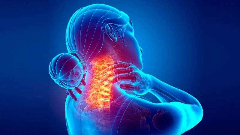
Spinal pathology

Lumbar spine surgery: disc herniation and stenosis

Minimally invasive endoscopic and microsurgical techniques for lumbar disc herniation, spinal canal stenosis, spondylolisthesis and degenerative disease. Fast recovery.

Lumbar spine surgery: disc herniation and stenosis

Lumbar spine surgery is the most common surgical treatment of the spine. Lumbar disc herniation is the leading cause of sciatica in adults (affecting 2% of the population each year); lumbar spinal canal stenosis is the leading cause of neurogenic claudication in people over 60. Current techniques are minimally invasive (microdiscectomy, endoscopy, mini-invasive TLIF) with recovery in days.
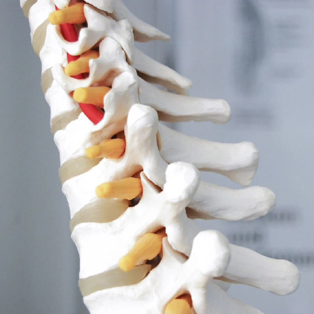
Lumbar spine and lumbar vertebrae
2%
annual incidence of lumbar disc herniation
90%
improve with conservative treatment within 6–12 weeks
<24h
typical hospital stay in lumbar endoscopic surgery

When should you see a neurosurgeon?

The most common symptoms are:

  • Sciatica: pain radiating down the leg following a dermatome (L4, L5 or S1)
  • Mechanical low back pain that worsens with bending or prolonged standing
  • Tingling or numbness in the leg or foot
  • Focal weakness (foot drop, quadriceps weakness)
  • Neurogenic claudication: leg pain or paraesthesia when walking that eases on bending forward (pathognomonic of spinal canal stenosis)
  • Sphincter dysfunction or saddle anaesthesia (cauda equina syndrome · EMERGENCY)
  • Disabling chronic low back pain with functional limitation
Urgent care: Cauda equina syndrome (loss of sphincter control, perineal anaesthesia, acute bilateral weakness) is a surgical emergency requiring surgery within the first 24–48h.

Techniques we use in the lumbar spine

Lumbar endoscopic surgery

Lumbar endoscopic discectomy

What it involves: removal of the herniated disc through a percutaneous approach.

  • Same-day discharge
  • Fast recovery

Indications: disc herniation.

Endoscopic decompression

What it involves: release of compressed nerve structures.

  • Minimal bone resection

Indications: stenosis.

Minimally invasive surgery (MIS)

Microdiscectomy

What it involves: removal of the herniation through a small incision.

  • Effective technique
  • Fast recovery

Indications: disc herniation.

Tubular decompression

What it involves: access to the canal using dilators.

  • Preserves the muscles

Indications: stenosis.

MIS lumbar fusion surgery

TLIF (posterior transforaminal)

What it involves: access to the disc through the foramen for decompression and fusion.

  • Less neural manipulation
  • Fusion through a single approach

Indications: spondylolisthesis, stenosis with instability.

PLIF (posterior bilateral)

What it involves: posterior access to the intervertebral disc.

  • High stability
  • Solid fusion

Indications: advanced degeneration, instability.

LLIF / XLIF (lateral)

What it involves: lateral access to the intervertebral disc.

  • Deformity correction
  • Muscle preservation

Indications: scoliosis, multilevel disease.

ALIF (anterior)

What it involves: direct anterior access to the intervertebral disc.

  • Restoration of lordosis
  • Large implants

Indications: disc degeneration, deformity.

Hybrid anterolateral techniques + posterior fusion

What it involves: combination of anterior or lateral approaches with percutaneous posterior fixation.

  • Maximum stability
  • Global correction

Indications: complex cases, deformity, revision surgery.

Lumbar percutaneous instrumentation

What it involves: screw placement using minimally invasive techniques.

  • Less surgical trauma
  • Faster recovery

Indications: lumbar fusion.

Lumbar robotic surgery

Lumbar robotic surgery (Alaya)

What it involves: guided implant placement using a robotic system.

  • Millimetre precision
  • Greater safety

Indications: multilevel surgery, complex cases.

How we approach each case

Every lumbar condition is assessed with a lumbar MRI and, in selected cases, a CT scan for bone assessment, standing radiographs for dynamic instability or an EMG to confirm the level of radiculopathy. Conservative treatment (physiotherapy, NSAIDs, guided epidural or selective nerve root injections) is the first line for 6–8 weeks. Surgery is indicated in: progressive motor deficit, refractory radicular pain lasting >6 weeks, cauda equina syndrome (urgent), spinal canal stenosis with disabling claudication or progressive instability.
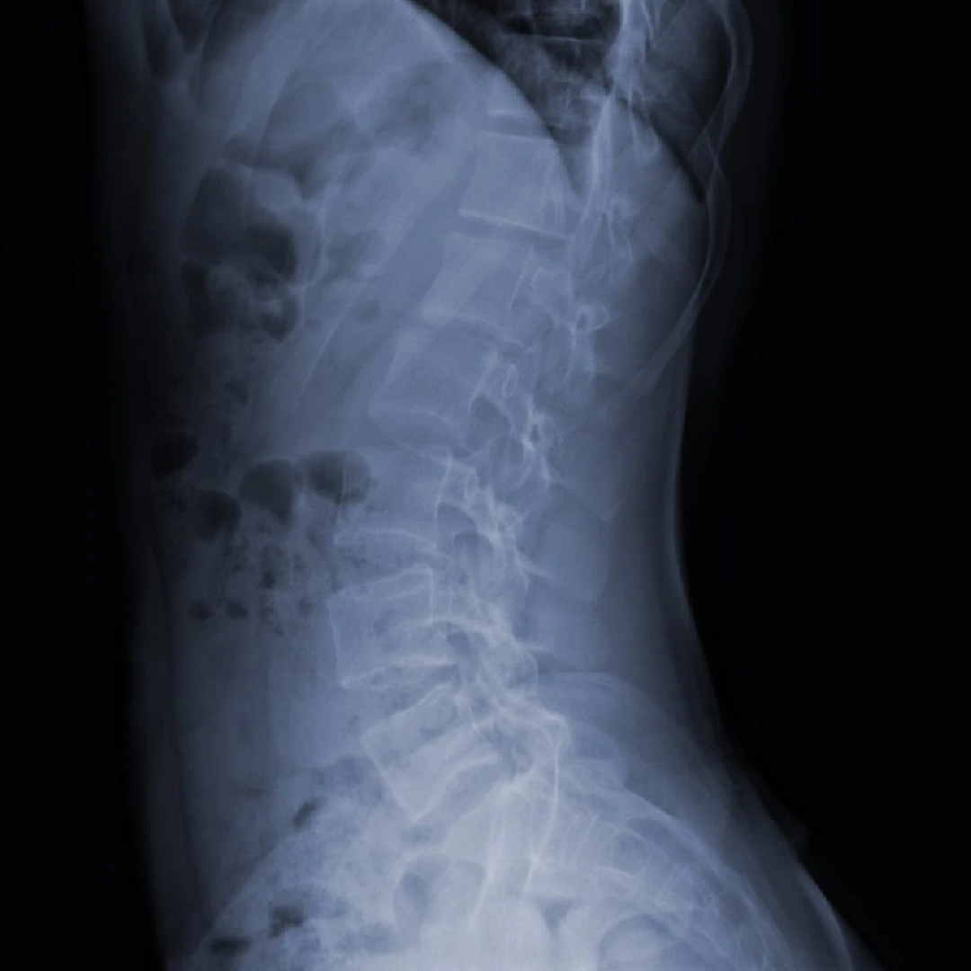
Lumbar spine surgery protocol

How we organise each case

  1. 1 · Preoperative assessment

    Lumbar MRI (standard), CT if bone assessment is required, standing radiographs for dynamic instability, EMG in cases with a deficit to confirm the nerve root level. Conservative treatment for 6–8 weeks before considering non-urgent surgery.

  2. 2 · Surgery

    Minimally invasive technique whenever possible: tubular microdiscectomy, endoscopy or mini-invasive TLIF. In fusions, ALAYA robotic assistance for planning and precise placement of pedicle screws.

  3. 3 · Immediate postoperative period

    Mobilisation within a few hours. Radiographic check at one month. In endoscopy/microdiscectomy, discharge usually within <24h; in fusion, 2–4 days.

  4. 4 · Discharge and follow-up

    Progressive physiotherapy from the first week. Return to office work in 2–3 weeks, physical work in 6–8. Follow-up with radiographs at 6 weeks, 3, 6 and 12 months.


Neurosurgeons who treat it

Dr. Jorge Torales

Dr. Jorge Torales

Neurosurgeon · Skull base and spinal endoscopy
View profile →
Dr. Abel Ferrés Pijoan

Dr. Abel Ferrés Pijoan

Neurosurgeon · Spine, neuro-oncology and skull base
View profile →
Dr. Pedro Roldán Ramos

Dr. Pedro Roldán Ramos

Neurosurgeon · Spine, functional and brain surgery
View profile →
Dr. Jhon A. Hoyos Castro

Dr. Jhon A. Hoyos Castro

Neurosurgeon · Spine, peripheral nerve and skull base
View profile →
Dra. María Elena Filadoro

Dra. María Elena Filadoro

Neurosurgeon · Functional, epilepsy and spine
View profile →
Dra. María Noelia Sosa Echeverría

Dra. M. Noelia Sosa Echeverría

Neurosurgeon · Functional and stereotactic surgery
View profile →
Dra. Giulia Guizzardi

Dra. Giulia Guizzardi

Neurosurgeon · Minimally invasive surgery and skull base
View profile →

Prognosis and recovery

Lumbar microdiscectomy achieves significant relief of radicular pain in 85–95% of selected patients. Endoscopic surgery achieves equivalent results with a faster recovery. In spinal canal stenosis, decompression improves walking capacity in 80% of patients at 2 years. In spondylolisthesis with instability, instrumented fusion achieves functional improvement in 75–85% of cases. The typical hospital stay ranges from 24h (endoscopy) to 3 days (fusion).

Answers to your questions

When should a lumbar disc herniation be operated on?
Most lumbar disc herniations improve with conservative treatment within 6–12 weeks. Surgery is indicated when there is persistent radicular pain lasting >6 weeks with functional impairment, progressive motor deficit, or cauda equina syndrome (an emergency: loss of sphincter control, saddle anaesthesia). The latter requires surgery within the first 24–48h.
What is lumbar microdiscectomy?
It is the most widely used surgical technique for lumbar disc herniation. It is performed through a small incision (2–3 cm), using a microscope or high-definition loupes, and only the portion of the disc compressing the nerve root is removed. The rest of the disc remains intact so that it keeps its function.
What is endoscopic spine surgery?
It is a minimally invasive surgical technique in which the disc is reached through a 7–8 mm endoscopic tube via a transforaminal or interlaminar approach. It reduces muscle damage, speeds up recovery and even allows same-day discharge in selected cases.
When is a lumbar fusion necessary?
Spinal fusion is indicated when there is vertebral instability: spondylolisthesis, degenerative disc disease with disabling mechanical pain, scoliosis, or after extensive decompressive surgery. Modern techniques (TLIF, LLIF, ALIF) are minimally invasive in most cases.
How long does recovery after lumbar spine surgery take?
After microdiscectomy or endoscopy: return to light activity within 1 week, office work in 2–3 weeks, physical work in 6–8 weeks. After fusion: recovery is more gradual, between 3 and 6 months until complete fusion. Physiotherapy begins in the first postoperative weeks.

What the clinical guidelines say

"Minimally invasive lumbar surgical techniques (microdiscectomy, endoscopy) are equivalent in effectiveness to traditional techniques, with advantages in postoperative pain and recovery time."
· North American Spine Society (NASS) · source
"Cauda equina syndrome is a neurosurgical emergency requiring decompression within the first 24–48 hours in order to maximise functional recovery."
· British Association of Spine Surgeons (BASS) · source

Scientific references consulted

Studies and clinical guidelines on which we base our approach to this condition.

  1. Weinstein JN. et al. Surgical versus nonoperative treatment for lumbar disc herniation (SPORT). N Engl J Med. 2006. View publication
  2. Lurie JD. et al. Long-term outcomes of lumbar spinal stenosis: 8-year results of the SPORT trial. Spine. 2015. View publication
  3. Forsth P. et al. A randomized, controlled trial of fusion surgery for lumbar spinal stenosis. N Engl J Med. 2016. View publication
  4. Qaseem A. et al. Noninvasive treatments for acute, subacute, and chronic low back pain (ACP guideline). Ann Intern Med. 2017. View publication
  5. Ruetten S. et al. Full-endoscopic interlaminar and transforaminal lumbar discectomy versus microsurgical technique. Spine. 2008. View publication
  6. North American Spine Society (NASS) Clinical Guidelines for Diagnosis and Treatment of Lumbar Disc Herniation with Radiculopathy. NASS. 2020. View publication

Related resources

Robot-assisted surgery with ALAYA

We use the ALAYA Robotic Assistant (Cyber Surgery) for the precise placement of pedicle screws in spinal fusion surgery, both open and minimally invasive.

ALAYA guides the trajectory of the surgical instruments through a patented kinematic tracking system that requires no optical navigation cameras: it avoids the classic line-of-sight problems and takes up less space in theatre. The patient's position is monitored in real time by a bone reference clamp attached directly to the tracking device, which improves the overall accuracy of the procedure.

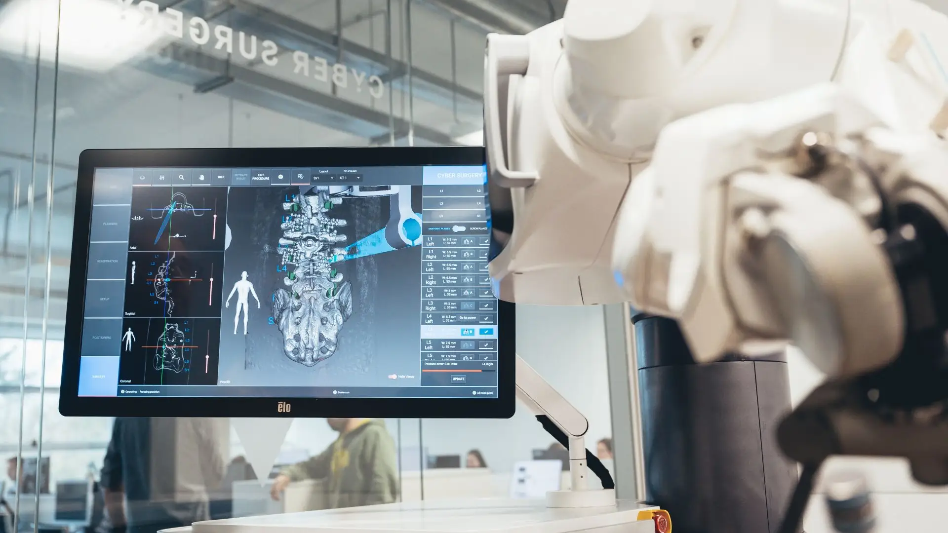
ALAYA robot by Cyber Surgery · ALAYA robot deployed in theatre during spinal fusion surgery.
ALAYA robot deployed in theatre during spinal fusion surgery.

What do we use it for?

  • Pedicle screw placement in the posterior thoracic and sacro-lumbar spine.
  • Spinal fusion surgery, both open and minimally invasive.
  • Intraoperative 3D planning using preoperative or intraoperative 2D/3D CT.
  • Implant-brand independence: compatible with any commercially available cannulated screw.
A real benefit for the patient: a reduction in mean hospital stay from 9.4 to 6.8 days documented with robotic assistance in spinal fusion, together with lower intraoperative radiation exposure for both patient and team.
ALAYA robotic assistant by Cyber Surgery in the operating theatre
Video: ALAYA Robotic Assistant (Cyber Surgery) in the operating theatre. Click to play.
Greater accuracy

Significantly more accurate pedicle screw placement than with conventional technique.

Minimally invasive

Less tissue damage, lower risk of infection and faster postoperative recovery.

Shorter operating time

An intuitive workflow and fast set-up that shorten the length of the procedure.

Lower radiation

The system minimises the number of image acquisitions needed during surgery.

Percutaneous discectomy with the Disc-FX system

Patient reviews

What our patients say

5,0 Average rating on Doctoralia · testimonials anonymised with consent
High-grade glioma

"I arrived with a glioma I had been told was inoperable. Here they reviewed it at the tumour board and offered me a plan. The surgery was a success. Four years on, I am well."

M
M. · 52 years old
Operated on in 2022
Cervical disc herniation

"For years I had a cervical disc herniation, operated on by another team with no improvement. Minimally invasive surgery gave me back my mobility and took the pain away."

A
A. · 48 years old
Revision after a first surgery
International patient

"I travelled from outside Spain. The team reviewed my case before I came and gave me a clear plan. Caring people and impeccable technique."
L
L. · 39 years old
Remote second opinion
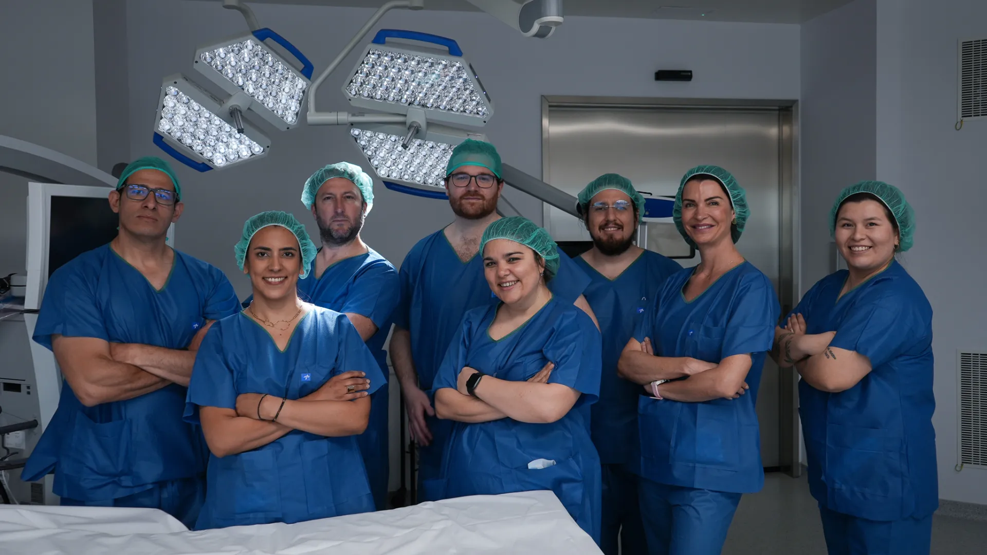
Medical team at Brain & Spine Barcelona: neurosurgeons, neurologists and nursing staff
A multidisciplinary team by your side. Neurosurgeons, neurologists and nursing staff working together on every case. Meet the whole team →
Request information

Lumbar spine consultation

Send us your lumbar MRI and your specialist's report. We will advise you on whether your case is suitable for conservative treatment, endoscopic surgery or microsurgery.

Add specialty of interest (optional)

We reply within 24-48 working hours.