Dr. Jorge Torales
Neurosurgeon · Skull base and spinal endoscopy
View profile
Microsurgical treatment of intramedullary and extramedullary pathology: spinal cord tumours, syringomyelia, spinal vascular malformations, and compressive cervical myelopathy. Continuous neurophysiological monitoring.
Spinal cord surgery treats lesions within the spinal cord or in close relationship to it. Intramedullary tumours (astrocytomas, ependymomas, haemangioblastomas) account for 5-10% of central nervous system tumours. Surgery requires high-definition microscopy, continuous neurophysiological monitoring, and specific expertise. The goal is maximum resection with functional preservation.
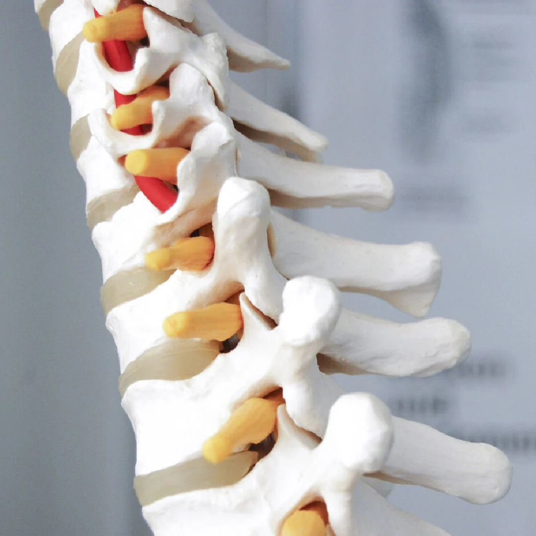
Symptoms depend on the spinal cord level affected:
The most common tumour in adults. A cleavage plane with the spinal cord allows complete resection in 90% of cases with functional preservation.
Infiltrative tumours, more difficult to resect completely. Goal: maximum safe resection with continuous monitoring.
Vascular tumours, sometimes associated with von Hippel-Lindau disease. En-bloc resection with spinal cord preservation.
When the cause is treatable (Chiari decompression), the syrinx usually resolves. In refractory cases, syringosubarachnoid or syringoperitoneal shunting.
Anterior approach (ACDF, corpectomy) or posterior approach (laminectomy, laminoplasty) depending on location and cause.
Dural fistulas and cavernomas. Microsurgical surgery or coordination with the endovascular team.
The workup includes contrast-enhanced spinal MRI (axial and sagittal sequences, T1/T2, contrast) and, where indicated, selective spinal angiography. All patients are assessed with a quantitative neurological scale (McCormick, JOA) preoperatively. Surgery is performed under high-definition microscopy with continuous neurophysiological monitoring (SSEP, MEP, EMG) and, where appropriate, intraoperative ultrasonography. Typical postoperative hospital stay is 5-10 days and includes early-onset neurological rehabilitation.
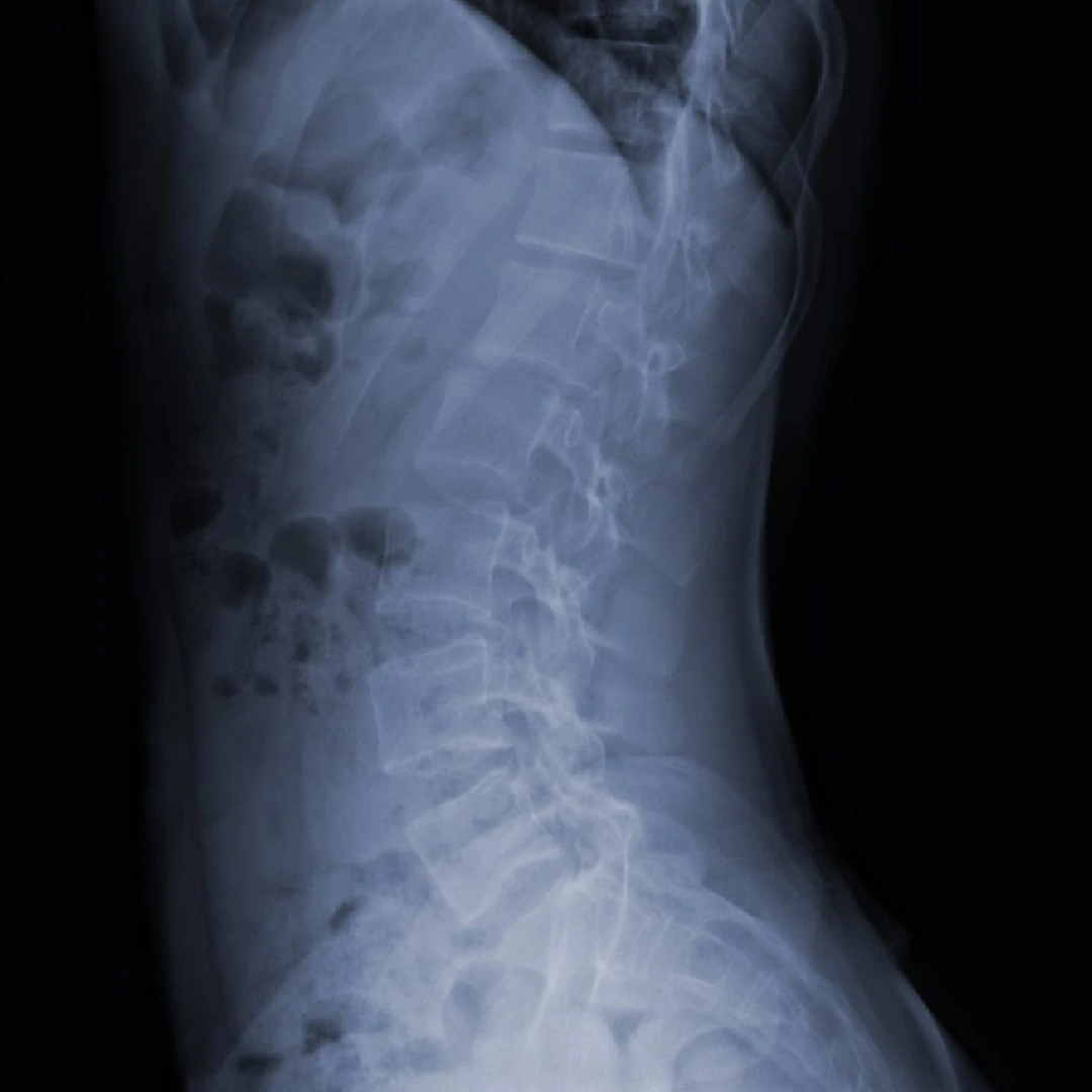
Gadolinium-enhanced spinal MRI with specific sequences, motor and somatosensory evoked potentials, functional assessment using ASIA and McCormick scales.
Microsurgery with continuous neurophysiological monitoring (MEPs, SSEPs, D-wave). Posterior, lateral, or anterior approaches according to the lesion. Ultrasonic aspirator (CUSA) and visualisation with intraoperative staining.
"From purely anatomical neurosurgery towards precision neurosurgery."
ICU 24-48h. Early integrated rehabilitation from day one. Serial functional assessment.
Discharge at 5-10 days with transition to a spinal rehabilitation unit. Follow-up MRI at 3, 6, and 12 months.
In spinal ependymomas, complete resection is achieved in 80-90% of cases with good long-term functional outcomes (survival >90% at 10 years). In astrocytomas, maximum safe resection prolongs survival, though recurrence is more frequent. Neurophysiological monitoring has reduced permanent new neurological deficit to below 5% at specialist centres. Postoperative recovery includes intensive neurological rehabilitation with functional follow-up for at least 12 months.
"Surgery for intramedullary tumours should be performed at specialist centres with full neurophysiological monitoring (SSEP, MEP) and specific microsurgical expertise."
"Total resection is the goal in well-defined spinal ependymomas and is associated with long-term disease-free survival exceeding 90%."
Studies and clinical guidelines on which we base our approach to this condition.
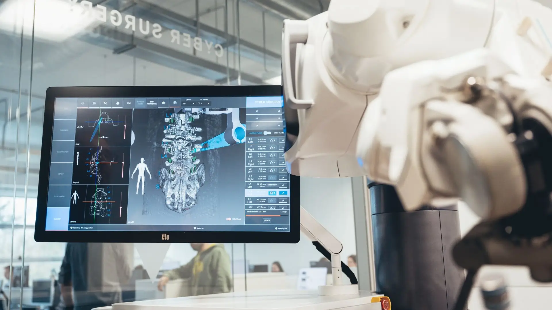
We use the ALAYA Robotic Assistant (Cyber Surgery) for the precise placement of pedicle screws in spinal fusion surgery, both open and minimally invasive.
ALAYA guides the trajectory of the surgical instruments through a patented kinematic tracking system that requires no optical navigation cameras: it avoids the classic line-of-sight problems and takes up less space in theatre. The patient's position is monitored in real time by a bone reference clamp attached directly to the tracking device, which improves the overall accuracy of the procedure.

Significantly more accurate pedicle screw placement than with conventional technique.
Less tissue damage, lower risk of infection and faster postoperative recovery.
An intuitive workflow and fast set-up that shorten the length of the procedure.
The system minimises the number of image acquisitions needed during surgery.
"I arrived with a glioma I had been told was inoperable. Here they reviewed it at the tumour board and offered me a plan. The surgery was a success. Four years on, I am well."
"For years I had a cervical disc herniation, operated on by another team with no improvement. Minimally invasive surgery gave me back my mobility and took the pain away."
"I travelled from outside Spain. The team reviewed my case before I came and gave me a clear plan. Caring people and impeccable technique."
Send us your contrast-enhanced spinal MRI and neurological report. We review the case in a multidisciplinary committee and respond with a personalised plan.