Dr. Jorge Torales
Neurosurgeon · Skull base and spinal endoscopy
View profile
Acoustic neuromas are benign tumours of the vestibular nerve. Skull base surgery allows preservation of the facial nerve and, in selected tumours, also hearing.
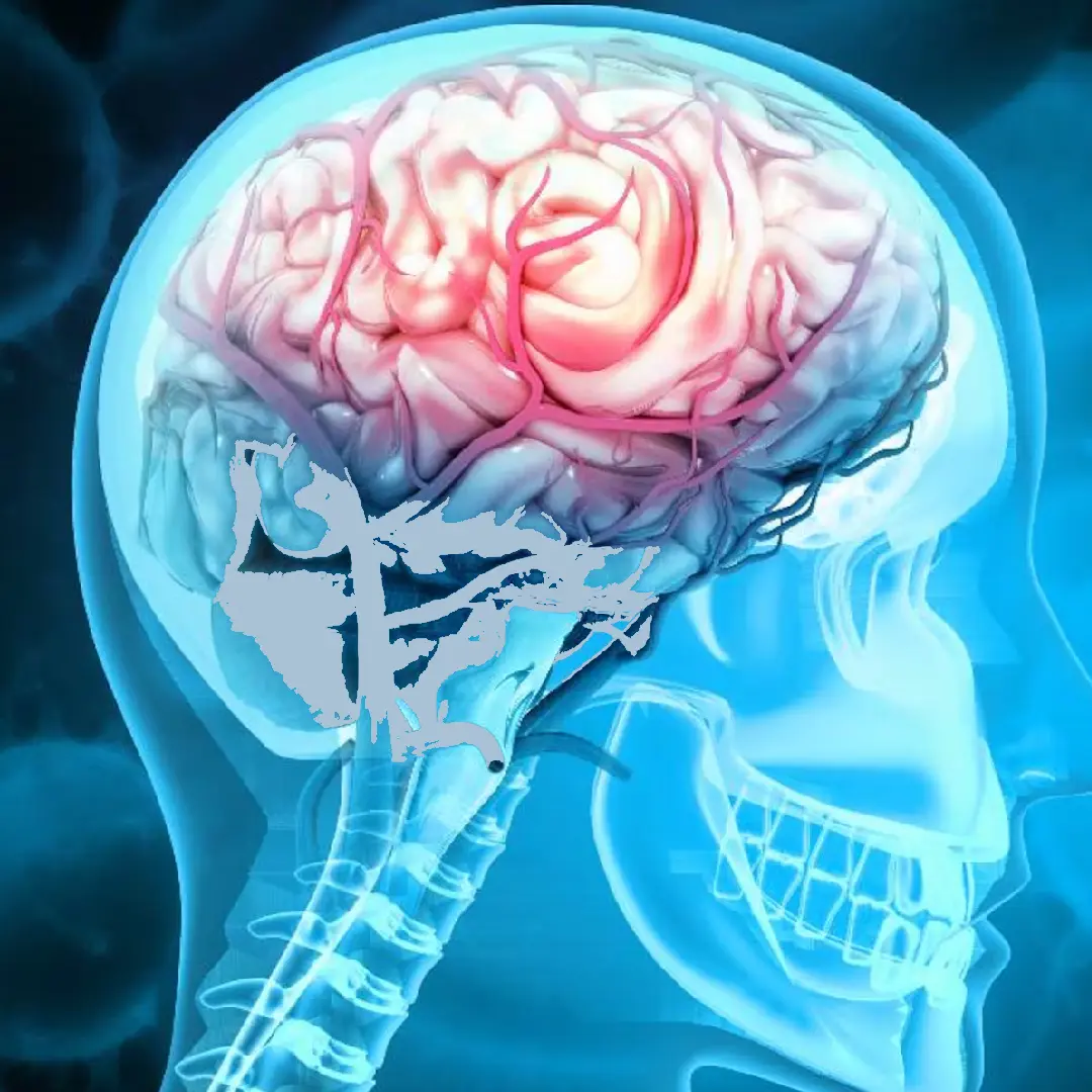
Acoustic neuromas, also known as vestibular schwannomas, are benign tumours that grow in the myelin sheath of the vestibular nerve. They account for 8% of intracranial tumours and 80% of cerebellopontine angle tumours. Initial symptoms are progressive unilateral hearing loss, tinnitus and unsteadiness. Treatment may be observation, radiosurgery or microsurgery depending on size and residual hearing.
Symptoms evolve slowly over years:
Technique of choice. It allows facial nerve preservation and, in tumours of 2 cm or less with useful preoperative hearing, also hearing preservation in 40-70% of cases.
Technique of choice in tumours with non-functional hearing. Better facial nerve preservation, at the cost of hearing.
Indicated in small intracanalicular tumours when hearing preservation is the primary goal.
Gamma Knife or CyberKnife for tumours <3 cm or as treatment for residual tumour. Tumour control of 90-95% at 10 years, but with a relevant side effect: most patients lose hearing within 2 years of follow-up. It is therefore not the first option when hearing preservation is a realistic goal.
In small tumours (<1.5 cm) that are asymptomatic or cause mild symptoms, especially in patients over 65. Annual MRI.
Continuous intraoperative EMG throughout surgery to identify and protect the facial nerve in real time.
Each case is studied with contrast-enhanced MRI (CISS/FIESTA sequences), pure-tone and speech audiometry, auditory brainstem-evoked potentials (ABR) and facial nerve assessment (House-Brackmann). The therapeutic decision is made on an individual basis considering size, residual hearing, patient age and preferences. When surgery is undertaken, continuous facial nerve monitoring (EMG) is used and, when appropriate, cochlear nerve monitoring as well. Reconstruction of the surgical defect uses autologous abdominal fat and bone cement to prevent CSF fistula.
It is not uncommon for acoustic neuroma to present with hydrocephalus due to obstruction of normal cerebrospinal fluid flow. In that case, the first step is treating the hydrocephalus: usually with a temporary external ventricular drain and, depending on the clinical course, consideration of a permanent drain (ventriculo-peritoneal shunt). Once hydrocephalus is controlled, neuroma surgery is planned.
MRI of the internal auditory canal with and without contrast, pure-tone and speech audiometry, auditory evoked potentials (ABR), vestibular assessment. Koos scale and Hannover classification.
Retrosigmoid, translabyrinthine or middle fossa approach depending on size and hearing preservation goal. Continuous facial and cochlear nerve monitoring. Microsurgery with functional preservation as the priority.
ICU for 24h. Postoperative MRI within 72h. Facial assessment with House-Brackmann scale. Follow-up audiometry.
Discharge after 5-7 days. Vestibular rehabilitation and, if facial weakness is present, specific physiotherapy. Annual MRI follow-up for at least 5 years.
Facial nerve preservation in specialist centres exceeds 95% in tumours <2 cm and 85% in large tumours. Even when postoperative facial palsy occurs, in most cases it is temporary and recovers progressively over the following months, including in large tumours. Surgical mortality is below 1%. Typical hospital stay is 5-7 days and return to work occurs between 6 and 8 weeks. Balance adapts progressively over 4-8 weeks (vestibular compensation).
On hearing: no technique can restore hearing that was already lost before surgery. The realistic goal is preserving existing hearing, not restoring it. In tumours <2 cm with useful preoperative hearing, approaches that spare the cochlear nerve achieve hearing preservation in 40-70% of cases.

"Treatment of vestibular schwannoma requires a multidisciplinary team comprising neurosurgery, otology and intraoperative neurophysiology. Functional outcomes are directly proportional to the volume and specialisation of the centre."
"Observation is a reasonable strategy in small tumours that are asymptomatic or cause only mild symptoms."
Studies and clinical guidelines on which we base our approach to this condition.
"I arrived with a glioma I had been told was inoperable. Here they reviewed it at the tumour board and offered me a plan. The surgery was a success. Four years on, I am well."
"For years I had a cervical disc herniation, operated on by another team with no improvement. Minimally invasive surgery gave me back my mobility and took the pain away."
"I travelled from outside Spain. The team reviewed my case before I came and gave me a clear plan. Caring people and impeccable technique."
Send us your MRI and audiometry. We review your case in our skull base board and advise on the strategy we recommend: observation, radiosurgery or surgery.