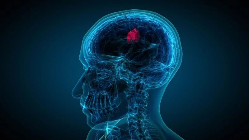
Brain tumour surgery
Maximum safe resection of brain tumours using neuronavigation, awake craniotomy and continuous neurophysiological monitoring to protect neurological functions.
A different approach for every tumour
In brain oncology no two tumours are alike. The surgical plan depends on the histological type, location, relationship with eloquent areas and the patient's condition. Each case is assessed by a multidisciplinary board before the strategy is decided.
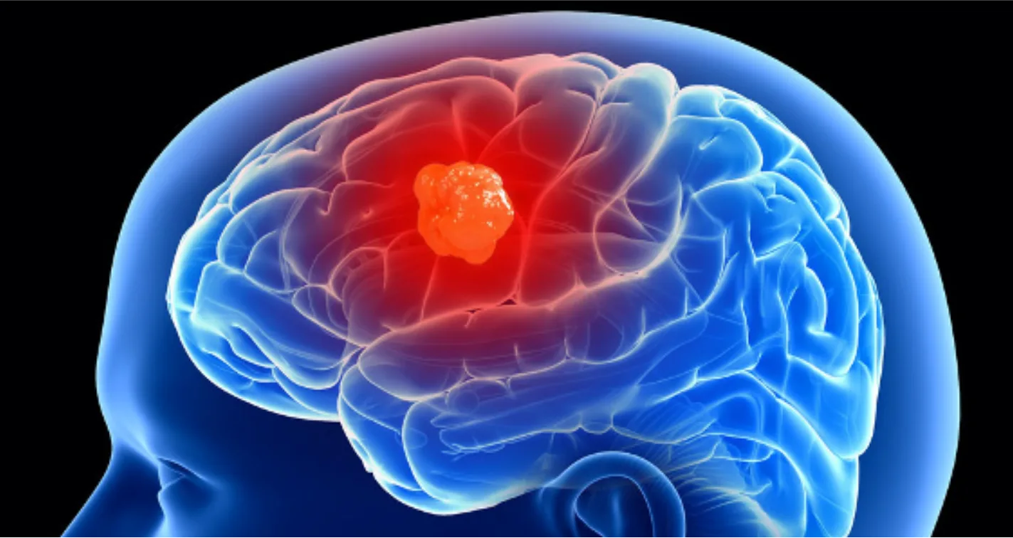
Symptoms of brain tumours
The symptoms of a brain tumour depend on its size, its rate of growth and, above all, its location. They may be caused by increased pressure within the skull or by direct involvement of a specific brain area.
General symptoms (due to raised intracranial pressure)
- Progressive headache, predominantly in the morning, worsening over time and with manoeuvres such as coughing or bending.
- Nausea and vomiting, sometimes without prior nausea ("sudden-onset vomiting").
- Drowsiness or slowing of the level of consciousness.
- Blurred or double vision and papilloedema on fundoscopy.
Focal symptoms (according to location)
- New-onset seizures (first seizure in an adult with no prior history).
- Focal neurological deficit: weakness or numbness on one side of the body.
- Speech disturbance (difficulty speaking or understanding).
- Visual disturbance or visual field defect.
- Unsteadiness, lack of coordination or gait disturbance (posterior fossa or cerebellar tumours).
Cognitive and behavioural changes
- Short-term memory loss and difficulty concentrating.
- Personality changes, apathy or irritability.
- Disorientation and slowed thinking.
Brain tumours we treat
Gliomas
Low- and high-grade. Maximum safe resection with neuronavigation, awake craniotomy and neurophysiological monitoring.
Meningiomas
Complete microsurgical resection with low recurrence rates. Preservation of adjacent vascular structures.
Acoustic neuromas
Skull base approaches with hearing and facial nerve preservation in selected tumours.
Maximum precision, preserved function
- Neuronavigation: high-resolution brain GPS to guide every millimetre.
- Awake craniotomy: real-time functional mapping in tumours within eloquent areas.
- Continuous neurophysiological monitoring: protection of speech, movement and cranial nerves.
- 4K/3D microscopy and intraoperative MRI when indicated.
- Multidisciplinary board: neurosurgery, neuro-oncology, neuroradiology and pathology working together.
MRI-guided laser ablation for brain tumours
Laser interstitial thermal therapy (LITT) has become established as an alternative or complement to open surgery in selected oncological scenarios. It does not replace maximum safe resection when that is feasible: it is one more tool, particularly useful when the lesion is deep, surrounded by eloquent areas, the patient is not a candidate for craniotomy, or when treating a recurrence without reopening a previously operated or irradiated field.
Glioblastoma and high-grade gliomas
Growing indication in recurrent GBM and in primary deep non-resectable cases. Modern series report an acceptable safety profile and survival data comparable to alternative rescue strategies4-8.
Brain metastases and radionecrosis
Particularly useful in recurrences after radiosurgery and in symptomatic radionecrosis refractory to corticosteroids. Ablation allows treatment of the lesion and, in many cases, histological distinction between tumour recurrence and necrosis9-11.
Deep or complex-location lesions
Insular, thalamo-mesencephalic or posterior fossa tumours where conventional microsurgical approach would carry high functional morbidity. The stereotactic trajectory reaches the target while minimising the lesioned corridor12,13.
Patient not suitable for craniotomy
Advanced age, comorbidity, history of multiple surgeries or prior irradiation. LITT offers a minimal-incision profile (4 mm), short stay and lower infection risk, with real-time thermal control via intraoperative MRI.

Scientific references (17)
Foundational and classic technical works
- Jethwa PR, Barrese JC, Gowda A, Shetty A, Danish SF. Magnetic resonance thermometry-guided laser-induced thermal therapy for intracranial neoplasms: initial experience. Neurosurgery. 2012;71(1 Suppl Operative):133-145. doi:10.1227/NEU.0b013e31826101d4.
- Hawasli AH, Bagade S, Shimony JS, Miller-Thomas M, Leuthardt EC. Magnetic resonance imaging-guided focused laser interstitial thermal therapy for intracranial lesions: single-institution series. Neurosurgery. 2013;73(6):1007-1017.
- Patel NV, Mian M, Stafford RJ, et al. Laser interstitial thermal therapy technology, physics of MRI thermometry, and technical considerations. Neurosurgery. 2016;79(Suppl 1):S8-S16.
Glioblastoma and high-grade gliomas
- Kamath AA, Friedman DD, Akbari SHA, et al. Glioblastoma treated with MRI-guided laser interstitial thermal therapy: safety, efficacy, and outcomes. Neurosurgery. 2019;84(4):836-843.
- de Groot JF, Kim AH, Prabhu S, et al. Efficacy of laser interstitial thermal therapy (LITT) for newly diagnosed and recurrent IDH wild-type glioblastoma. Neuro-Oncol Adv. 2022.
- Mortezaei A, et al. Laser interstitial thermal therapy for high-grade glioma. Neurosurg Focus. 2025.
- Pandey A, et al. Safety and efficacy of laser interstitial thermal therapy as upfront treatment for primary brain tumors. Cancers (Basel). 2024.
- Montemurro N, et al. Survival outcomes in recurrent glioblastoma treated with LITT: a systematic review. Clin Neurol Neurosurg. 2020.
Brain metastases and radionecrosis
- Srinivasan ES, Sankey EW, Grabowski MM, Chongsathidkiet P, Fecci PE. Laser interstitial thermal therapy for brain metastases. Neuro-Oncol Adv. 2021.
- Sujijantarat N, Hong CS, Owusu KA, et al. LITT versus bevacizumab for radiation necrosis in previously irradiated brain metastases. J Neurooncol. 2020.
- Chan M, et al. Efficacy of laser interstitial thermal therapy for biopsy-proven radiation necrosis. J Neurooncol. 2023.
Deep tumours and complex locations
- Vetkas A, et al. Laser interstitial thermal therapy for the treatment of insular lesions: a systematic review. Front Neurol. 2022.
- Ashraf O, et al. MR-guided LITT for posterior fossa neoplasms. J Neurooncol. 2020.
Complications and functional outcomes
- Sharma M, et al. Thermal injury to corticospinal tracts and postoperative motor deficits after LITT. Neurosurg Focus. 2016.
- Wilhelmy B, et al. Functional outcomes following LITT for recurrent high-grade glioma. J Neurooncol. 2024.
Modern reviews and state of the art
- Patel B, et al. Laser interstitial thermal therapy. Neuroimaging Clin N Am. 2020.
- O'Halloran PJ, et al. LITTing up gliomas, is the future bright? Curr Oncol Rep. 2023.
- Sriram S, et al. Updates on the role and efficacy of LITT in neuro-oncology. Adv Radiat Oncol. 2023.
Neurosurgeons in the oncological area


Dr. Pedro Roldán Ramos

Dr. Jorge Torales

Dr. Jhon A. Hoyos Castro

Dr. María Elena Filadoro

Dr. M. Noelia Sosa Echeverría

Dr. Giulia Guizzardi
Brain tumour resection in 60 seconds
Related resources
What our patients say
"I arrived with a glioma I had been told was inoperable. Here they reviewed it at the tumour board and offered me a plan. The surgery was a success. Four years on, I am well."
"For years I had a cervical disc herniation, operated on by another team with no improvement. Minimally invasive surgery gave me back my mobility and took the pain away."
"I travelled from outside Spain. The team reviewed my case before I came and gave me a clear plan. Caring people and impeccable technique."
Second opinion in 24-48h
We review your test results and give you a personalised plan before any travel.