Trauma surgery

Neurotrauma: treatment of traumatic brain injury (TBI)

Urgent surgical management and follow-up of traumatic brain injury: intracranial haematomas, cerebral contusions, skull fractures, and chronic sequelae. 24-hour immediate response.

Neurotrauma: treatment of traumatic brain injury

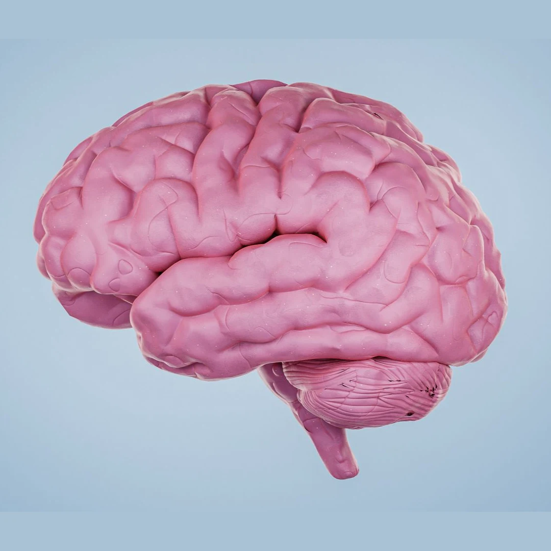
Neurotrauma encompasses injuries to the nervous system caused by trauma. Traumatic brain injury (TBI) is one of the leading causes of death in people under 45. Intracranial haematomas (extradural, subdural, intraparenchymal) may require urgent surgery. The time from injury to treatment is the single most important modifiable prognostic factor.
Neurotrauma: treatment of traumatic brain injury
1h
critical window in extradural haematoma for maximum recovery
40-60%
mortality of acute subdural haematoma without surgery
70-80%
functional recovery after timely surgery for chronic haematoma

When to consult a neurosurgeon?

After a head injury, warning signs include:

  • Loss of consciousness, even briefly, following the impact
  • Lucid interval followed by deterioration (classic sign of extradural haematoma)
  • Progressive headache not relieved by analgesics
  • Projectile vomiting, especially in children
  • Altered speech, vision, or movement
  • Confusion, irritability, or progressive drowsiness
  • Pupil asymmetry or inability to be roused
  • Post-traumatic seizures
Urgent attention: Any head injury with loss of consciousness, vomiting, progressive headache, or neurological deterioration requires an urgent CT scan. Time is the critical factor.

Techniques we use

Acute subdural haematoma evacuation

Urgent surgery via wide craniotomy. Prognosis is directly proportional to the speed of evacuation (golden rule: within the first 4 hours).

Chronic subdural haematoma drainage

Burr hole under local or general anaesthesia. The liquefied haematoma is drained through two holes. Excellent prognosis with discharge in 2-3 days.

Extradural haematoma surgery

Urgent craniotomy. Prognosis is excellent if surgery is performed before cerebral herniation · many young patients achieve full functional recovery.

Decompressive craniectomy

Removal of a large bone flap to allow expansion of cerebral oedema. Indicated for refractory raised intracranial pressure. Reconstruction (cranioplasty) is performed 2-6 months later.

ICP monitoring

Intraventricular or intraparenchymal catheter for continuous intracranial pressure measurement. Guides treatment in severe TBI (Glasgow ≤8).

Depressed skull fracture repair

Elevation of the depressed bone fragment when there is neurological compromise or dural laceration. The dura is repaired and the skull reconstructed.

How we approach each case

Every patient with severe TBI (Glasgow ≤8) is admitted to the neurocritical care unit with ICP monitoring, invasive arterial blood pressure monitoring, and cerebral oximetry. The Brain Trauma Foundation protocol guides management: maintaining ICP below 22 mmHg, cerebral perfusion pressure 60-70 mmHg, normoxia, and normocapnia. When intracranial hypertension is refractory, medical treatment is escalated (deep sedation, osmotherapy, hypothermia) and decompressive craniectomy is considered as a last resort. Post-discharge follow-up includes neurorehabilitation, neuropsychology, and radiological review.

How we organise each case

  1. 1 · Pre-operative assessment

    Stabilisation of the polytrauma patient (ABCDE), urgent CT scan, Glasgow Coma Scale assessment, and Marshall / Rotterdam CT scoring.

  2. 2 · Surgery

    Immediate surgical evacuation of haematomas with mass effect. Decompressive craniectomy when ICP exceeds thresholds refractory to medical treatment.

  3. 3 · Immediate post-operative care

    Neurocritical care unit with continuous ICP, CPP, and, where indicated, cerebral oxygenation monitoring (PtiO₂). Application of the Brain Trauma Foundation protocol.

  4. 4 · Discharge and follow-up

    Variable according to severity. Comprehensive neurological rehabilitation coordinated with neuropsychology and speech therapy. Surgical follow-up and deferred skull reconstruction if craniectomy was performed.
Neurosurgeons who treat this condition

Dr. Jorge Torales

Dr. Jorge Torales

Neurosurgeon · Skull base and spinal endoscopy
View profile →
Dr. Abel Ferrés Pijoan

Dr. Abel Ferrés Pijoan

Neurosurgeon · Spine, neuro-oncology, and skull base
View profile →
Dr. Pedro Roldán Ramos

Dr. Pedro Roldán Ramos

Neurosurgeon · Spine, functional, and brain surgery
View profile →
Dr. Jhon A. Hoyos Castro

Dr. Jhon A. Hoyos Castro

Neurosurgeon · Spine, peripheral nerve, and skull base
View profile →
Dra. María Elena Filadoro

Dra. María Elena Filadoro

Neurosurgeon · Functional, epilepsy, and spine
View profile →
Dra. María Noelia Sosa Echeverría

Dra. M. Noelia Sosa Echeverría

Neurosurgeon · Functional and stereotaxy
View profile →
Dra. Giulia Guizzardi

Dra. Giulia Guizzardi

Neurosurgeon · Minimally invasive and skull base
View profile →

Prognosis and recovery

Prognosis depends on initial severity (Glasgow), age, associated injuries, and speed of treatment. In extradural haematoma with early surgery, full recovery occurs in 70-80% of otherwise healthy young patients. In acute subdural haematoma, mortality remains high despite treatment (30-60%). In chronic subdural haematoma, 80-90% of patients regain their previous level of function. Severe TBI may leave motor, cognitive, or behavioural sequelae in a significant proportion of patients; early neurological rehabilitation improves functional outcome.
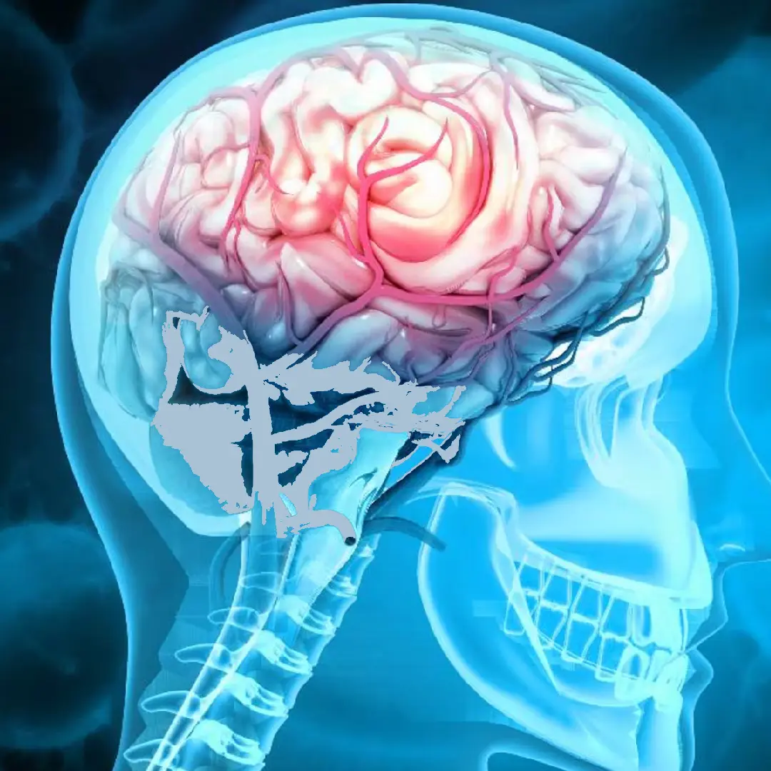
Prognosis and recovery after traumatic brain injury

Answers to your questions

What is an acute subdural haematoma?
It is a collection of blood between the dura mater and the arachnoid following a traumatic brain injury. It results from rupture of bridging veins and carries a high mortality (40-60%) if not treated urgently. Surgical evacuation via craniotomy is required within the first few hours.
What about a chronic subdural haematoma?
This is a slowly evolving blood collection (over weeks), common in older patients, those on anticoagulants, or following minor trauma. It is treated with a small burr hole and drainage of the liquefied haematoma. The prognosis is excellent and patients are typically discharged in 2-3 days.
When is surgery indicated for a traumatic brain injury?
Surgery is indicated for haematomas with significant mass effect (midline shift >10 mm or volume >30 cc), expanding lesions with neurological deterioration, refractory raised intracranial pressure, or depressed skull fractures with neurological compromise.
What is intracranial pressure (ICP) monitoring?
It is a fine catheter that continuously measures ICP following severe TBI. It allows early detection of raised intracranial pressure and guides treatment (cerebrospinal fluid drainage, osmotherapy, deep sedation, or decompressive craniectomy if refractory).
Can someone fully recover after a TBI?
Recovery depends on initial severity (Glasgow Coma Scale), the anatomical injury, and the speed of treatment. Mild TBI usually results in full recovery. Severe TBI (GCS 3-8) may leave motor, cognitive, or behavioural sequelae. Early neurological rehabilitation improves functional outcome.

What the clinical guidelines say

"The time from injury to surgical treatment is the single most important modifiable prognostic factor in acute intracranial haematomas."
· Brain Trauma Foundation · source
"Intracranial pressure monitoring in severe TBI (Glasgow ≤8) is the standard of care for guiding treatment and improving outcomes."
· Society of Critical Care Medicine · source

Scientific references consulted

Studies and clinical guidelines on which our approach to this condition is based.

  1. Carney N. et al. Guidelines for the Management of Severe Traumatic Brain Injury, 4th Edition. Neurosurgery. 2017. View publication
  2. Cooper DJ. et al. Decompressive craniectomy in diffuse traumatic brain injury (DECRA). N Engl J Med. 2011. View publication
  3. Hutchinson PJ. et al. Trial of decompressive craniectomy for traumatic intracranial hypertension (RESCUEicp). N Engl J Med. 2016. View publication
  4. Teasdale G, Jennett B. Assessment of coma and impaired consciousness: a practical scale (GCS). Lancet. 1974. View publication
  5. Maas AIR. et al. Prediction of outcome in traumatic brain injury with computed tomographic characteristics: Rotterdam score. Neurosurgery. 2005. View publication
Patient reviews

What our patients say

5,0 Average rating on Doctoralia · testimonials anonymised with consent
High-grade glioma

"I arrived with a glioma I had been told was inoperable. Here they reviewed it at the tumour board and offered me a plan. The surgery was a success. Four years on, I am well."

M
M. · 52 years old
Operated on in 2022
Cervical disc herniation

"For years I had a cervical disc herniation, operated on by another team with no improvement. Minimally invasive surgery gave me back my mobility and took the pain away."

A
A. · 48 years old
Revision after a first surgery
International patient

"I travelled from outside Spain. The team reviewed my case before I came and gave me a clear plan. Caring people and impeccable technique."
L
L. · 39 years old
Remote second opinion
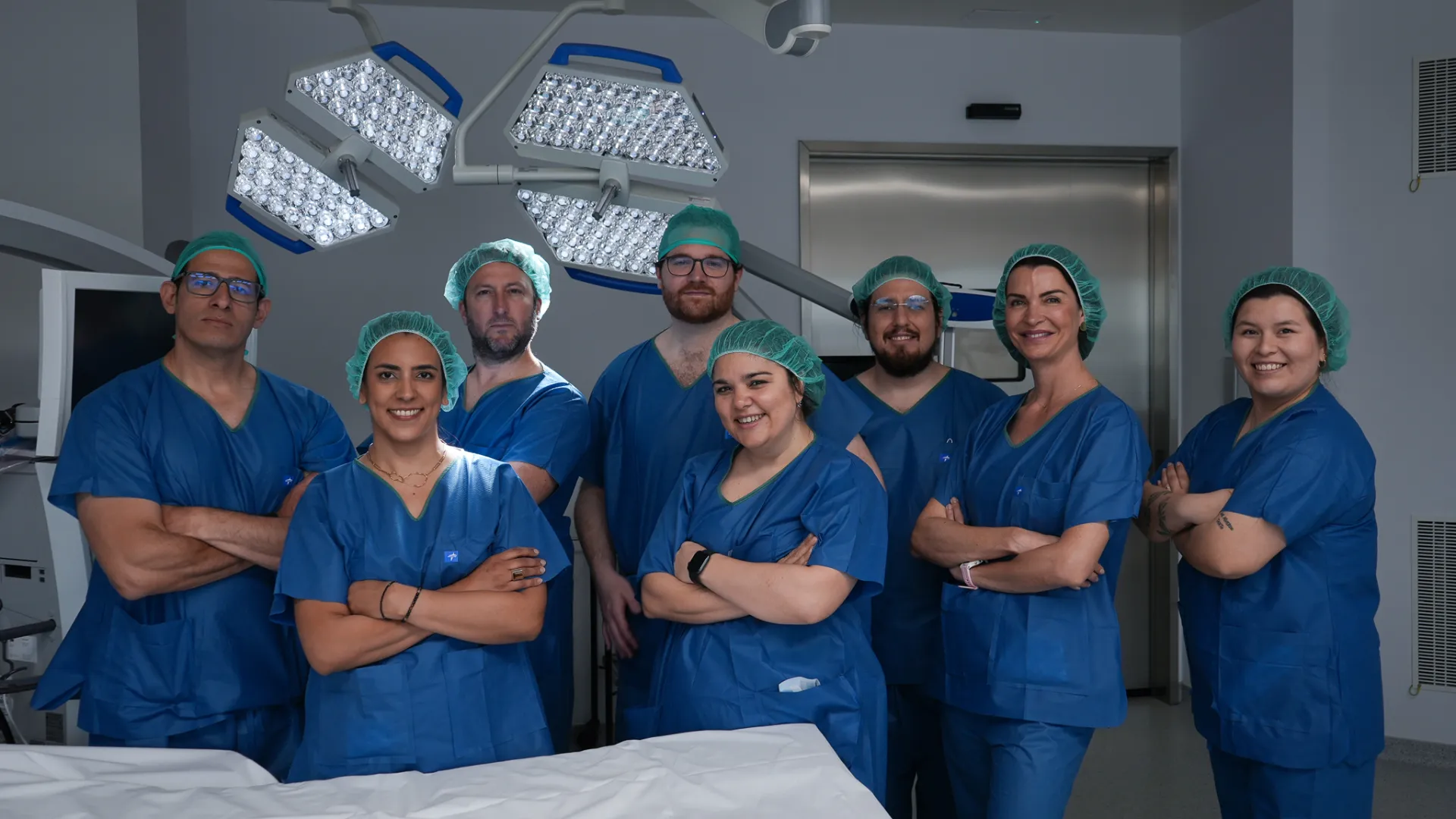
Medical team at Brain & Spine Barcelona: neurosurgeons, neurologists and nursing staff
A multidisciplinary team by your side. Neurosurgeons, neurologists and nursing staff working together on every case. Meet the whole team →
Request information

Assessment after head injury

If you have sustained a TBI and would like a review of sequelae or post-operative progress, please send us your study results and clinical report. Response within 24-48 hours.

Add specialty of interest (optional)

We reply within 24-48 working hours.