Dr. Abel Ferrés Pijoan
Neurosurgeon · Spine, neuro-oncology and skull base
View profile →
Microsurgical and minimally invasive treatment of cervical pathology: disc herniation, spinal stenosis, spondylotic myelopathy, traumatic pathology. Motion preservation with arthroplasty where possible.
Cervical spine surgery treats conditions that cause radicular pain or myelopathy due to neural compression. Cervical disc herniation is the most common cause in younger adults; cervical spinal stenosis and spondylotic myelopathy are more typical from the age of 50. The most commonly used techniques are ACDF (discectomy and fusion) and, in selected cases, cervical arthroplasty with a disc prosthesis that preserves motion.
The most common symptoms include:
What it involves: anterior approach to remove the affected disc and decompress the spinal cord or nerve roots, followed by fusion.
Indications: cervical disc herniation, myelopathy, stenosis.
What it involves: fusion of two or more vertebrae using implants.
Indications: instability, advanced degeneration.
What it involves: expansion of the spinal canal without completely removing posterior structures.
Indications: multi-level myelopathy.
What it involves: posterior canal decompression via a minimally invasive approach.
Indications: cervical stenosis.
What it involves: treatment of herniations or compressions via a percutaneous endoscopic approach.
Indications: selected disc herniation, foraminal stenosis.
What it involves: placement of screws and rods for vertebral stabilisation.
Indications: trauma, tumours, instability.
What it involves: robotic assistance for cervical screw placement.
Indications: complex surgery, revision procedures.
Every patient with cervicobrachialgia is assessed with a cervical MRI and, when required, a CT scan for bony study. Suspected myelopathy is evaluated using the mJOA scale and electromyography. Initial treatment is conservative (physiotherapy, NSAIDs, guided injections) for 6-8 weeks before considering surgery. In progressive myelopathy, surgery is indicated early to prevent permanent spinal cord damage. The choice between ACDF, arthroplasty and posterior approach is individualised according to age, number of levels, morphology and cervical alignment.
Cervical MRI, CT for bony study, dynamic flexion-extension radiographs, neurological assessment with mJOA and Nurick scales for myelopathy.
Anterior approach (ACDF, arthroplasty) or posterior approach (laminoplasty, laminectomy with fixation) according to pathology. Neurophysiological monitoring in myelopathy.
Early mobilisation with soft cervical collar for 1-3 weeks. Radiographs at 6 weeks, 3 and 6 months.
Discharge within 24-48h after ACDF or arthroplasty, 3-5 days after posterior surgery. Physiotherapy from week 4.




ACDF for cervical disc herniation with radiculopathy achieves resolution of radicular pain in 85-90% of patients. Cervical arthroplasty has demonstrated non-inferiority to fusion in long-term follow-up, with advantages in motion preservation and reduced adjacent segment disease. In cervical myelopathy, early surgery stabilises or improves function in 70-80% of cases. Hospital stay is 1-3 days and return to work 3-6 weeks (office) or 3 months (physical work).
"Surgery should be considered in progressive symptomatic cervical myelopathy to halt neurological deterioration and improve function."
"Cervical arthroplasty is a reasonable alternative to fusion in selected patients with cervical disc disease without significant facet arthritis."
Studies and clinical guidelines on which we base our approach to this condition.
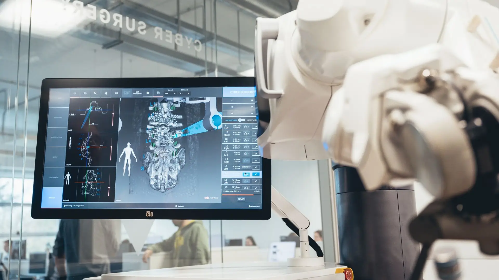
We use the ALAYA Robotic Assistant (Cyber Surgery) for the precise placement of pedicle screws in spinal fusion surgery, both open and minimally invasive.
ALAYA guides the trajectory of the surgical instruments through a patented kinematic tracking system that requires no optical navigation cameras: it avoids the classic line-of-sight problems and takes up less space in theatre. The patient's position is monitored in real time by a bone reference clamp attached directly to the tracking device, which improves the overall accuracy of the procedure.

Significantly more accurate pedicle screw placement than with conventional technique.
Less tissue damage, lower risk of infection and faster postoperative recovery.
An intuitive workflow and fast set-up that shorten the length of the procedure.
The system minimises the number of image acquisitions needed during surgery.
"I arrived with a glioma I had been told was inoperable. Here they reviewed it at the tumour board and offered me a plan. The surgery was a success. Four years on, I am well."
"For years I had a cervical disc herniation, operated on by another team with no improvement. Minimally invasive surgery gave me back my mobility and took the pain away."
"I travelled from outside Spain. The team reviewed my case before I came and gave me a clear plan. Caring people and impeccable technique."
Send us your cervical MRI and, if available, your electromyography. We will give you a clear opinion on whether your case is a candidate for conservative or surgical treatment.