Cerebrospinal fluid

Hydrocephalus and Chiari malformation

Comprehensive treatment of cerebrospinal fluid disorders in adults and children: communicating and obstructive hydrocephalus, adult normal-pressure hydrocephalus, Chiari malformation, and arachnoid cysts.

Hydrocephalus and Chiari malformation

Hydrocephalus is the abnormal accumulation of cerebrospinal fluid (CSF) in the cerebral ventricles, causing their dilation and raised intracranial pressure. It may be obstructive (due to a block in CSF flow) or communicating (due to impaired absorption). Chiari malformation type I (descent of the cerebellar tonsils >5 mm) can cause headaches, cerebellar symptoms, and syringomyelia. Both conditions require personalised neurosurgical assessment.
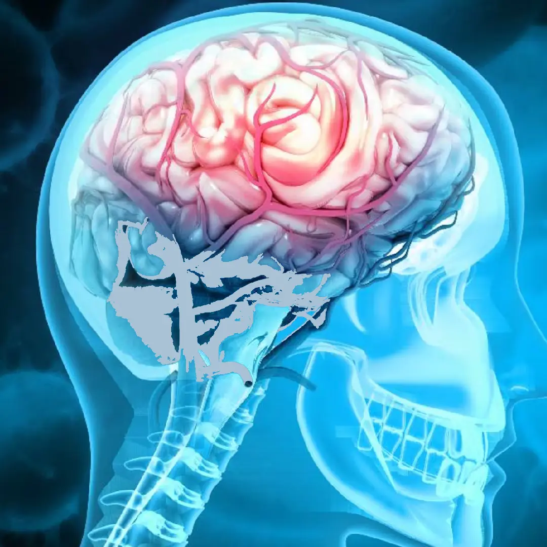
MRI and anatomy of cerebrospinal fluid
1/1000
prevalence of hydrocephalus in children
70%
ETV success rate in obstructive hydrocephalus
>80%
symptomatic improvement after surgery for symptomatic Chiari

When to consult a neurosurgeon?

Symptoms depend on age and cause:

  • Progressive headache, typically worse in the morning, improving on standing
  • Rapidly increasing head circumference in infants (cardinal sign)
  • Gait disturbance and loss of balance (normal-pressure hydrocephalus)
  • New-onset urinary incontinence in adults
  • Progressive cognitive decline (Hakim-Adams triad)
  • Occipital headache worsening with Valsalva or coughing (typical Chiari)
  • Cerebellar symptoms, dysarthria, or dysphagia in Chiari
  • Pain, weakness, or sensory loss due to associated syringomyelia
Urgent attention: Reduced level of consciousness, persistent vomiting, or pupil changes are emergencies requiring an urgent CT scan.

Techniques we use

Endoscopic third ventriculostomy (ETV)

Treatment of choice for obstructive hydrocephalus. An opening is made in the floor of the third ventricle for direct drainage into the subarachnoid space. Avoids implanted hardware.

Ventriculoperitoneal shunt

Shunt system for communicating hydrocephalus or failed ETV. Programmable valves adjusted non-invasively. Long-term follow-up is required.

Ventriculoatrial shunt

Alternative when peritoneal shunting is not feasible (abdominal adhesions, ascites). Direct drainage into the right atrium.

Craniovertebral decompression

Treatment of choice for symptomatic Chiari type I. Suboccipital craniectomy plus C1 laminectomy, with or without duraplasty, to increase the space and allow free CSF flow.

Endoscopic arachnoid cyst resection

Fenestration or resection of symptomatic cysts causing neurological compression or raised intracranial pressure.

ICP monitoring

In selected cases to confirm the diagnosis of normal-pressure hydrocephalus or pseudotumour cerebri by 24-72-hour continuous recording.

How we approach each case

Assessment includes brain and craniovertebral MRI with CSF flow sequences (phase-contrast cine MRI) to evaluate CSF dynamics. In adult normal-pressure hydrocephalus we perform a tap test (lumbar puncture with drainage of 40-60 ml) to predict the response to shunting. ETV is considered for all obstructive hydrocephalus with favourable anatomy (aqueductal stenosis, third-ventricle tumours). Shunts use the latest-generation programmable valves allowing non-invasive adjustments. In Chiari, surgery is indicated in cases with significant symptoms, documented progression, or associated syringomyelia.

How we organise each case

  1. 1 · Pre-operative assessment

    Brain and cervical MRI (including cine MRI to study CSF flow), lumbar infusion test or tap test for adult hydrocephalus, ophthalmological assessment if raised intracranial pressure is suspected.

  2. 2 · Surgery

    For shunts: programmable valve with anti-siphon device. For ETV: neuroendoscopy with perforation of the third ventricle floor. For Chiari: posterior fossa decompression with duraplasty when indicated.

  3. 3 · Immediate post-operative care

    CT or MRI review. Serial clinical assessment (gait, continence, headache). Valve pressure adjustment in programmable shunts.

  4. 4 · Discharge and follow-up

    Discharge after 3-5 days. Follow-up MRI at one month and at 6-12 months. Patient education on signs of shunt malfunction.

Neurosurgeons who treat this condition

Dr. Pedro Roldán Ramos

Dr. Pedro Roldán Ramos

Neurosurgeon · Spine, functional, and brain surgery
View profile →
Dr. Jorge Torales

Dr. Jorge Torales

Neurosurgeon · Skull base and spinal endoscopy
View profile →
Dr. Abel Ferrés Pijoan

Dr. Abel Ferrés Pijoan

Neurosurgeon · Spine, neuro-oncology, and skull base
View profile →
Dr. Jhon A. Hoyos Castro

Dr. Jhon A. Hoyos Castro

Neurosurgeon · Spine, peripheral nerve, and skull base
View profile →
Dra. María Elena Filadoro

Dra. María Elena Filadoro

Neurosurgeon · Functional, epilepsy, and spine
View profile →
Dra. María Noelia Sosa Echeverría

Dra. M. Noelia Sosa Echeverría

Neurosurgeon · Functional and stereotaxy
View profile →
Dra. Giulia Guizzardi

Dra. Giulia Guizzardi

Neurosurgeon · Minimally invasive and skull base
View profile →

Prognosis and recovery

ETV achieves a 70-80% success rate in obstructive hydrocephalus and avoids the complications associated with shunts. Modern shunts have failure rates of 10-15% in the first year (infection, malfunction). Chiari surgery in selected patients achieves symptomatic improvement in over 80% of cases and stabilises or reduces syringomyelia in the majority. Typical hospital stays are 2-4 days after ETV, 3-5 days after shunting, and 4-6 days after Chiari decompression.

Answers to your questions

What is hydrocephalus in adults?
It is an excess of cerebrospinal fluid (CSF) within the cerebral ventricles, causing their dilation and raised pressure. In adults it causes headache, gait disturbance, urinary incontinence, and cognitive decline (the Hakim-Adams triad in normal-pressure hydrocephalus).
What is endoscopic third ventriculostomy (ETV)?
It is a minimally invasive technique that creates an opening in the floor of the third ventricle to allow CSF to drain directly into the subarachnoid space, bypassing the obstruction. It is the treatment of choice for obstructive hydrocephalus and does not require a shunt implant.
When is a shunt indicated?
A ventriculoperitoneal shunt is indicated for communicating hydrocephalus, adult normal-pressure hydrocephalus, or when ETV has failed. Modern shunts are programmable, adjusted non-invasively by radiofrequency according to the required drainage pressure.
What is Chiari malformation and how is it treated?
It is the descent of the cerebellar tonsils below the foramen magnum. Type I in adults is associated with occipital headaches, neck pain, and sometimes syringomyelia. Surgical treatment (craniovertebral decompression) is indicated when there are significant symptoms or associated syringomyelia.
How long is the hospital stay after surgery?
After ETV: 2-4 days. After shunt implantation: 3-5 days. After craniovertebral decompression for Chiari: 4-6 days. Post-operative follow-up includes a control MRI and clinical assessment at 1, 3, and 12 months.

What the clinical guidelines say

"Endoscopic third ventriculostomy is the first therapeutic option in selected obstructive hydrocephalus, especially in cases of aqueductal stenosis."
· European Association of Neurosurgical Societies (EANS) · source
"The surgical indication for Chiari malformation type I should be based on the presence of clear clinical symptoms or associated syringomyelia, not solely on radiological criteria."
· Sociedad Española de Neurocirugía (SENEC) · source

Scientific references consulted

Studies and clinical guidelines on which our approach to this condition is based.

  1. Kulkarni AV. et al. Outcomes of endoscopic third ventriculostomy in children. Lancet. 2017. View publication
  2. Relkin N. et al. Diagnosing idiopathic normal-pressure hydrocephalus. Neurosurgery. 2005. View publication
  3. Milhorat TH. et al. Chiari I malformation redefined: clinical and radiographic findings for 364 symptomatic patients. Neurosurgery. 1999. View publication
  4. Friedman DI. et al. Revised diagnostic criteria for the pseudotumor cerebri syndrome. Neurology. 2013. View publication
  5. Rekate HL. A consensus on the classification of hydrocephalus. Childs Nerv Syst. 2011. View publication

Related resources

Patient reviews

What our patients say

5,0 Average rating on Doctoralia · testimonials anonymised with consent
High-grade glioma

"I arrived with a glioma I had been told was inoperable. Here they reviewed it at the tumour board and offered me a plan. The surgery was a success. Four years on, I am well."

M
M. · 52 years old
Operated on in 2022
Cervical disc herniation

"For years I had a cervical disc herniation, operated on by another team with no improvement. Minimally invasive surgery gave me back my mobility and took the pain away."

A
A. · 48 years old
Revision after a first surgery
International patient

"I travelled from outside Spain. The team reviewed my case before I came and gave me a clear plan. Caring people and impeccable technique."
L
L. · 39 years old
Remote second opinion
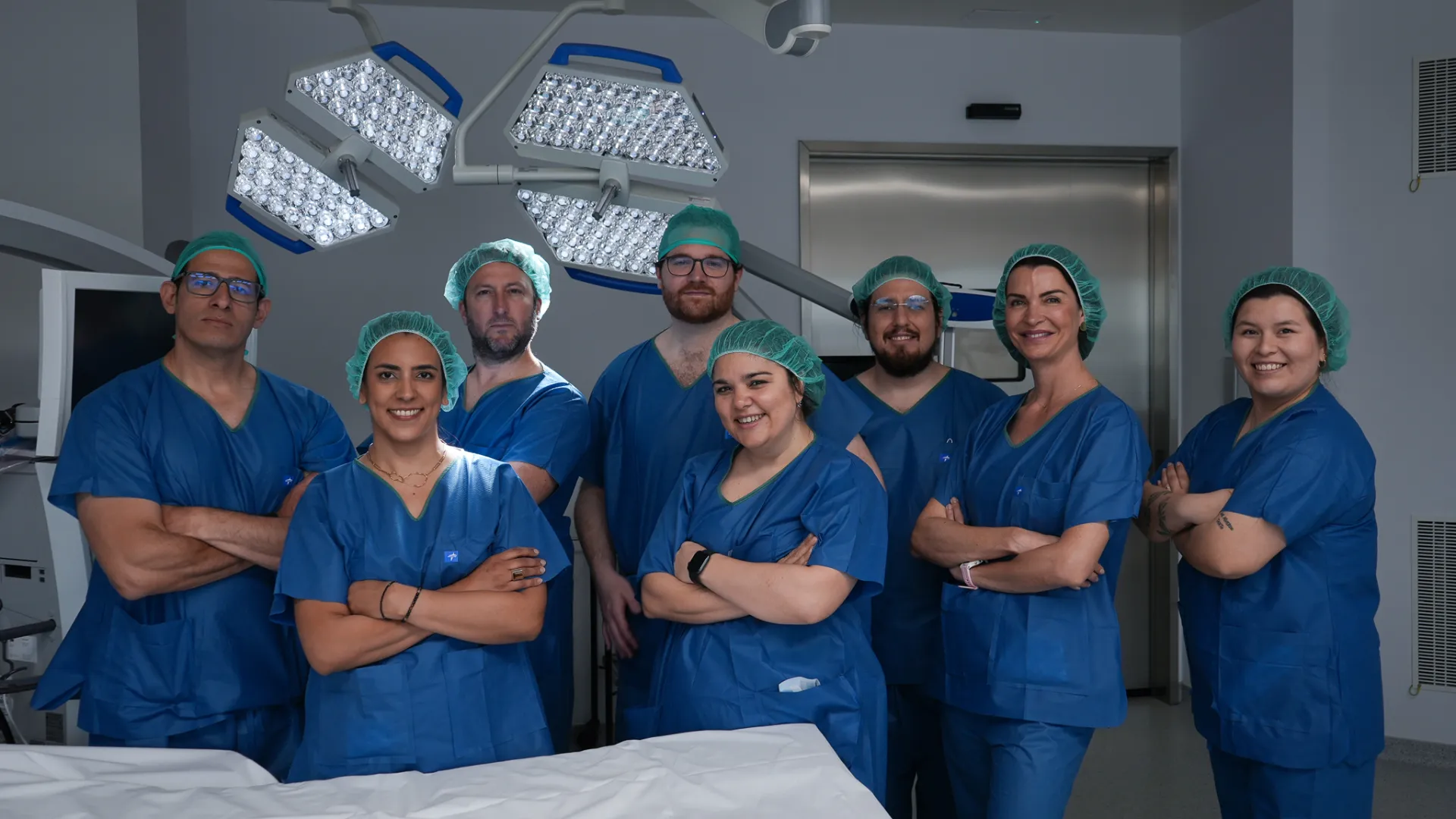
Medical team at Brain & Spine Barcelona: neurosurgeons, neurologists and nursing staff
A multidisciplinary team by your side. Neurosurgeons, neurologists and nursing staff working together on every case. Meet the whole team →
Request information

Enquiry about hydrocephalus or Chiari malformation

Send us your MRI scan (including CSF flow sequences if available) and we will assess whether surgical treatment is indicated for your case.

Add specialty of interest (optional)

We reply within 24-48 working hours.