
Spinal pathology

Thoracic spine surgery

Surgical treatment of thoracic vertebral pathology: thoracic disc herniations, vertebral tumours (primary and metastatic), thoracolumbar fractures and deformities (kyphosis, scoliosis).

Thoracic spine surgery

Thoracic spine surgery treats pathologies of the 12 thoracic vertebrae, a zone with complex anatomy due to its relationship with the rib cage, aorta, lungs and spinal cord. Thoracic disc herniations are uncommon (<1% of all herniations) but technically complex. Vertebral tumours (mainly metastases) and osteoporotic fractures are the most common reasons for surgery in this region.
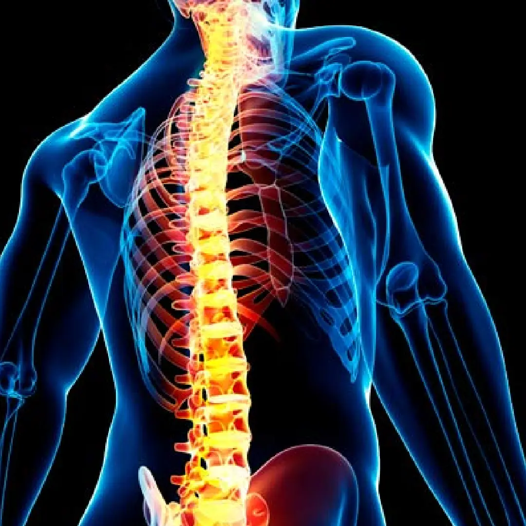
Thoracic vertebral spine
<1%
of disc herniations are thoracic
70%
of vertebral metastases are located in the thoracic spine
80%
pain improvement after kyphoplasty for osteoporotic fractures

When to see a neurosurgeon?

Symptoms vary according to cause:

  • Mechanical or referred dorsal pain in a thoracic band pattern
  • Intercostal radicular pain (well localised in the affected dermatome)
  • Leg weakness or gait disturbance (thoracic myelopathy)
  • Sensory disturbance below the level of the lesion
  • Bladder or bowel dysfunction in significant spinal cord compression
  • Intense focal pain following trauma (fracture)
  • Progressive visible deformity (hump) in kyphosis or scoliosis
Urgent attention: Rapid onset of paraparesis or loss of sensation in the legs requires urgent MRI: it may indicate acute spinal cord compression syndrome.

Techniques we use in thoracic spine surgery

Vertebroplasty

What it involves: injection of cement into the fractured vertebra.

  • Rapid pain relief
  • Percutaneous procedure

Indications: osteoporotic fractures.

Kyphoplasty

What it involves: insertion of a balloon to restore vertebral height before cement injection.

  • Deformity correction
  • Pain reduction

Indications: compression fractures.

MIS thoracic laminectomy

What it involves: decompression of the spinal canal via a minimally invasive approach.

  • Less tissue disruption
  • Faster recovery

Indications: thoracic stenosis, spinal cord compression.

Thoracic endoscopic surgery

What it involves: percutaneous approach to treat disc or compressive pathology.

  • Less tissue damage

Indications: selected thoracic herniations.

Percutaneous thoracic fusion

What it involves: stabilisation using screws placed in a minimally invasive fashion.

  • Less blood loss
  • Faster recovery

Indications: fractures, instability.

Robotic thoracic surgery (Alaya)

What it involves: robotic assistance for instrumentation placement.

  • High precision
  • Enhanced safety

Indications: tumours, deformities, complex surgery.

How we approach each case

The workup includes a complete thoracic MRI, CT scan for bony study and, when indicated, bone scintigraphy or PET-CT in suspected tumour. In vertebral metastases we apply the SINS (Spine Instability Neoplastic Score) and the Tokuhashi scale for the surgical decision. Fractures are evaluated using the TLICS classification. The approach is chosen according to the location of pathology (central, paramedian, lateral) and patient condition. In metastases with life expectancy >3 months and neurological deficit, surgery improves function and quality of life.
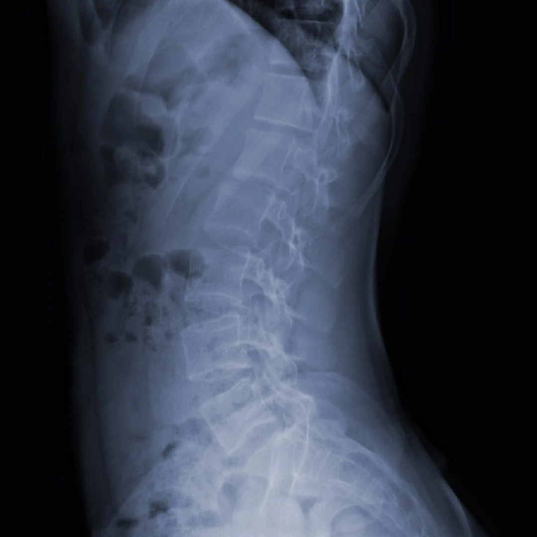
Thoracic spine surgery protocol

How we organise each case

  1. 1 · Preoperative workup

    Thoracic MRI with and without contrast, CT for bony study, bone densitometry for osteoporotic fractures, full oncological workup for metastases.

  2. 2 · Surgery

    Open or minimally invasive techniques according to lesion. For metastases, NOMS algorithm (Neurologic, Oncologic, Mechanical, Systemic). ALAYA robotic assistance for thoracic pedicle screws.

  3. 3 · Immediate postoperative period

    Early mobilisation. After vertebroplasty, discharge within 24h. After major procedures, progressive rehabilitation.

  4. 4 · Discharge and follow-up

    Discharge 1-7 days depending on procedure. Radiographic follow-up at 6 weeks and coordination with oncology where appropriate.


Neurosurgeons who treat this condition

Dr. Pedro Roldán Ramos

Dr. Pedro Roldán Ramos

Neurosurgeon · Spine, functional and brain surgery
View profile →
Dr. Jorge Torales

Dr. Jorge Torales

Neurosurgeon · Skull base and spinal endoscopy
View profile →
Dr. Abel Ferrés Pijoan

Dr. Abel Ferrés Pijoan

Neurosurgeon · Spine, neuro-oncology and skull base
View profile →
Dr. Jhon A. Hoyos Castro

Dr. Jhon A. Hoyos Castro

Neurosurgeon · Spine, peripheral nerve and skull base
View profile →
Dra. María Elena Filadoro

Dr. María Elena Filadoro

Neurosurgeon · Functional, epilepsy and spine
View profile →
Dra. María Noelia Sosa Echeverría

Dr. M. Noelia Sosa Echeverría

Neurosurgeon · Functional and stereotaxy
View profile →
Dra. Giulia Guizzardi

Dr. Giulia Guizzardi

Neurosurgeon · Minimally invasive and skull base
View profile →

Prognosis and recovery

Surgery for thoracic disc herniation achieves resolution of radicular pain in 75-85% and neurological improvement in the majority of patients operated for progressive thoracic myelopathy. Kyphoplasty for osteoporotic fracture relieves pain in 80% of patients within the first 48 hours. In vertebral metastases with spinal cord compression, early surgery combined with radiotherapy preserves the ability to walk in 70% of patients operated before losing ambulation.

Answers to your questions

Is thoracic disc herniation common?
No. Thoracic disc herniations represent less than 1% of all vertebral disc herniations because the thoracic spine is stabilised by the rib cage. When they occur they can be technically more complex as the spinal cord is in direct contact with the disc.
How are thoracic herniations operated on?
Several approaches exist: transthoracic (thoracotomy), costotransversectomy, lateral extracavitary approach, or thoracic endoscopic approach. The choice depends on disc location (central, paramedian or lateral) and the surgeon's preference.
What vertebral tumours are treated in the thoracic spine?
Both primary tumours (rare) and vertebral metastases (most commonly from lung, breast and prostate cancer). Treatment combines surgery (stabilisation, decompression, en-bloc resection where viable) with oncology, radiotherapy and systemic treatments.
When is surgery indicated for a thoracolumbar fracture?
Surgery is indicated for unstable fractures (compromise of all 3 Denis columns, kyphotic angulation >30°, wedging >50%) or in the presence of neurological deficit. Fixation can be percutaneous (minimally invasive) or open depending on the need for decompression.
What are kyphoplasty and vertebroplasty?
They are percutaneous techniques to treat painful osteoporotic vertebral fractures. Bone cement is injected into the vertebral body through a needle to stabilise it and relieve pain. Kyphoplasty adds the use of a balloon that restores some vertebral height before injecting the cement.

What clinical guidelines say

"Decompressive surgery combined with radiotherapy is superior to radiotherapy alone in vertebral metastases with spinal cord compression in patients with a reasonable life expectancy."
· Patchell RA et al., Lancet 2005 · source
"Vertebroplasty and kyphoplasty techniques are effective in relieving pain from osteoporotic fractures refractory to medical treatment."
· North American Spine Society (NASS) · source

Scientific references consulted

Studies and clinical guidelines on which we base our approach to this condition.

  1. Laufer I. et al. The NOMS framework: approach to the treatment of spinal metastatic tumors. Oncologist. 2013. View publication
  2. Klazen CAH. et al. Vertebroplasty versus conservative treatment in acute osteoporotic vertebral compression fractures (VERTOS II). Lancet. 2010. View publication
  3. Vaccaro AR. et al. AOSpine Thoracolumbar Spine Injury Classification System. Eur Spine J. 2013. View publication
  4. Patchell RA. et al. Direct decompressive surgical resection in the treatment of spinal cord compression caused by metastatic cancer. Lancet. 2005. View publication
  5. Fehlings MG. et al. Spinal cord injury evaluation and management guidelines (AOSpine). Global Spine J. 2017. View publication

Related resources

Robot-assisted surgery with ALAYA

We use the ALAYA Robotic Assistant (Cyber Surgery) for the precise placement of pedicle screws in spinal fusion surgery, both open and minimally invasive.

ALAYA guides the trajectory of the surgical instruments through a patented kinematic tracking system that requires no optical navigation cameras: it avoids the classic line-of-sight problems and takes up less space in theatre. The patient's position is monitored in real time by a bone reference clamp attached directly to the tracking device, which improves the overall accuracy of the procedure.

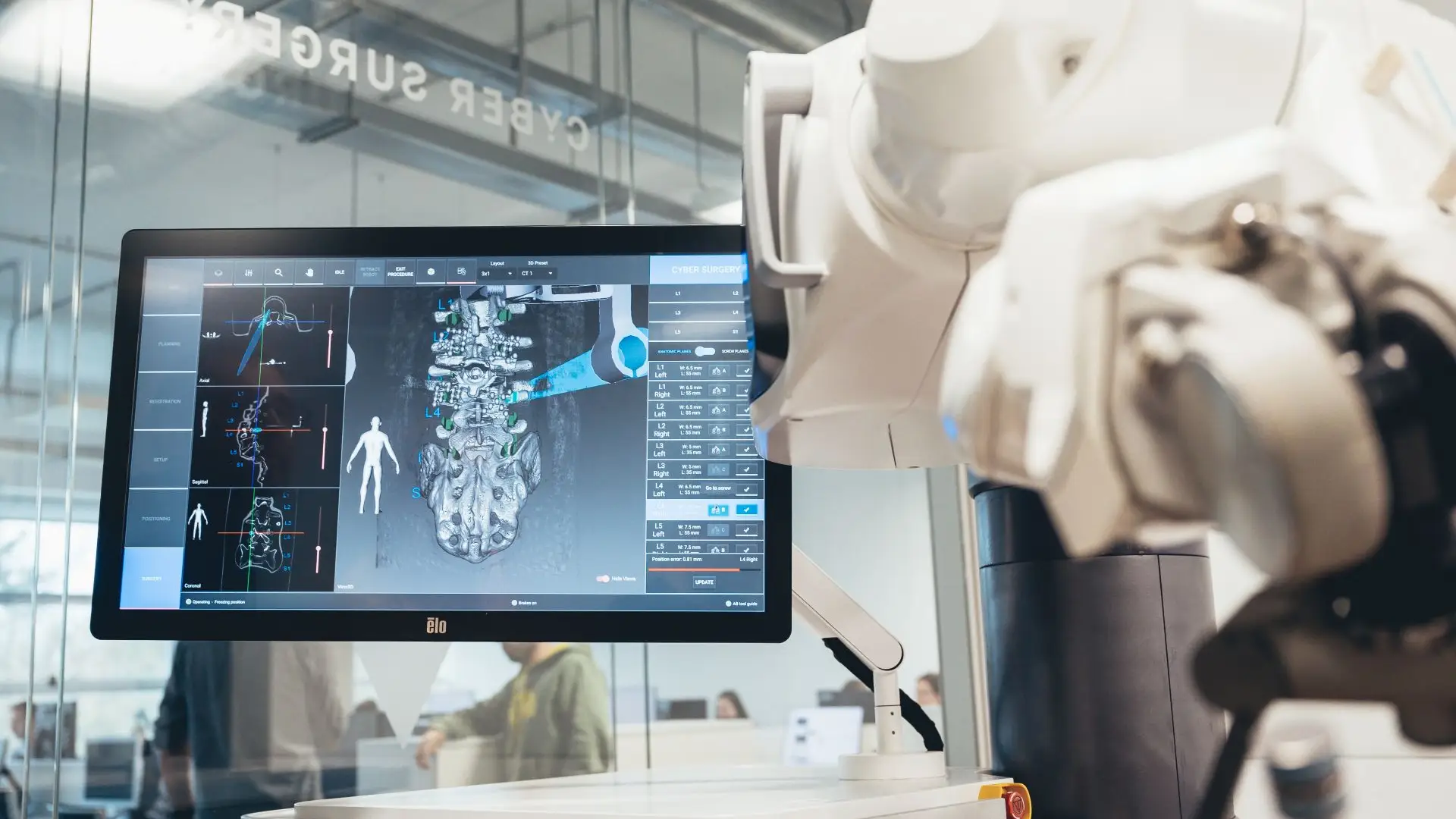
ALAYA robot by Cyber Surgery · ALAYA robot deployed in theatre during spinal fusion surgery.
ALAYA robot deployed in theatre during spinal fusion surgery.

What do we use it for?

  • Pedicle screw placement in the posterior thoracic and sacro-lumbar spine.
  • Spinal fusion surgery, both open and minimally invasive.
  • Intraoperative 3D planning using preoperative or intraoperative 2D/3D CT.
  • Implant-brand independence: compatible with any commercially available cannulated screw.
A real benefit for the patient: a reduction in mean hospital stay from 9.4 to 6.8 days documented with robotic assistance in spinal fusion, together with lower intraoperative radiation exposure for both patient and team.
ALAYA robotic assistant by Cyber Surgery in the operating theatre
Video: ALAYA Robotic Assistant (Cyber Surgery) in the operating theatre. Click to play.
Greater accuracy

Significantly more accurate pedicle screw placement than with conventional technique.

Minimally invasive

Less tissue damage, lower risk of infection and faster postoperative recovery.

Shorter operating time

An intuitive workflow and fast set-up that shorten the length of the procedure.

Lower radiation

The system minimises the number of image acquisitions needed during surgery.

Patient reviews

What our patients say

5,0 Average rating on Doctoralia · testimonials anonymised with consent
High-grade glioma

"I arrived with a glioma I had been told was inoperable. Here they reviewed it at the tumour board and offered me a plan. The surgery was a success. Four years on, I am well."

M
M. · 52 years old
Operated on in 2022
Cervical disc herniation

"For years I had a cervical disc herniation, operated on by another team with no improvement. Minimally invasive surgery gave me back my mobility and took the pain away."

A
A. · 48 years old
Revision after a first surgery
International patient

"I travelled from outside Spain. The team reviewed my case before I came and gave me a clear plan. Caring people and impeccable technique."
L
L. · 39 years old
Remote second opinion
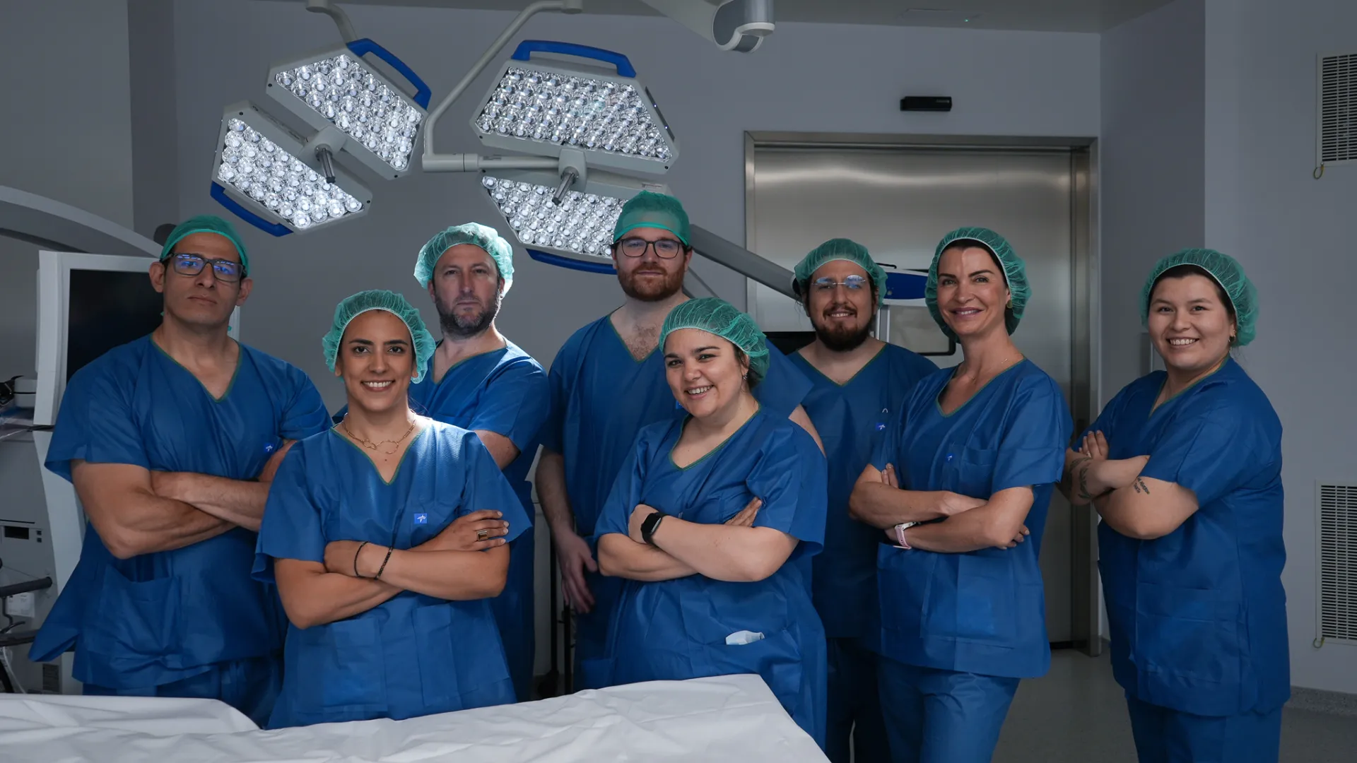
Medical team at Brain & Spine Barcelona: neurosurgeons, neurologists and nursing staff
A multidisciplinary team by your side. Neurosurgeons, neurologists and nursing staff working together on every case. Meet the whole team →
Request information

Thoracic spine consultation

Send us your thoracic MRI and CT scan if you have one. Thoracic herniations and pathologies in this region are technically complex · we will advise you on the best approach.

Add specialty of interest (optional)

We reply within 24-48 working hours.