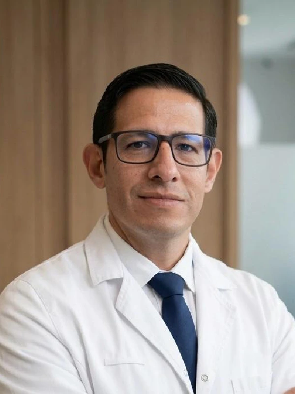
Dr. Jorge Torales
Neurosurgeon · Skull base and spinal endoscopy
View profile
Meningiomas are the most common primary brain tumours in adults and are generally benign. Complete resection surgery to Simpson grade I-II offers cure rates above 90% for grade I meningiomas.
Meningiomas are tumours that arise from the arachnoid cells of the meninges (the membranes covering the brain and spinal cord). They account for 38% of primary brain tumours and are generally benign (WHO grade I in 80% of cases). Surgery is the treatment of choice when they are symptomatic, and Simpson grade I-II resection is associated with recurrence rates below 10% at 10 years.
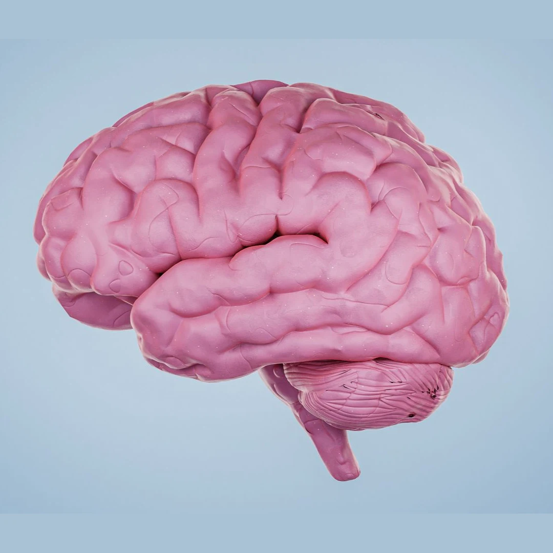
Many meningiomas are asymptomatic and discovered incidentally on imaging. When symptoms occur they typically include:
The technique of choice. Includes the tumour, the involved dura and 1 cm of adjacent bone. 4K/3D microscopy to preserve vessels and cranial nerves.
For sphenoid wing, clinoidal, petroclival or tentorial meningiomas. Pterional, orbitozygomatic, retrosigmoid or transpetrosal approaches depending on location.
Minimally invasive technique for meningiomas of the anterior cranial fossa and the clivus. Access via a transsphenoidal route (through the nose), with no brain retraction, no external scar and shorter recovery times.
In highly vascular meningiomas (especially convexity or falcine). Reduces intraoperative bleeding.
Gamma Knife or CyberKnife for residual tumour, recurrences or patients who are not surgical candidates. Effective for lesions under 3 cm.
For small asymptomatic meningiomas in older patients. MRI monitoring every 6-12 months initially.
Every patient with a meningioma is assessed with contrast-enhanced MRI and MR angiography when vascular involvement is suspected. In highly vascular meningiomas, diagnostic angiography and embolisation 24-48 hours before surgery are considered. We plan the approach using 3D preoperative modelling for complex locations. Surgery is performed with neurophysiological monitoring for meningiomas affecting cranial nerves or eloquent areas. Follow-up includes MRI at 3 months, 1 year and then annually for 5 years.
Brain MRI with gadolinium plus MR angiography to assess blood supply. Preoperative embolisation for hypervascular tumours. Ophthalmological and audiological assessment where the location requires it.
Approach planned according to location (convexity, skull base, parasagittal). Intraoperative neurophysiological monitoring. Microsurgery with a Simpson grade I-II resection target.
ICU for 24 hours in most cases. Postoperative MRI within 72 hours. Histopathology with WHO grading determines the follow-up schedule.
Discharge between 3 and 6 days depending on location. MRI at 3 months and annually for WHO grade I meningiomas. Complementary radiosurgery if resection is incomplete or grade II-III.
Complete Simpson grade I-II resection of grade I meningiomas is associated with cure rates above 90% at 10 years. Surgical mortality for convexity meningiomas is below 1% at high-volume centres. Atypical meningiomas (grade II) carry a higher recurrence risk (30-40% at 5 years) and usually require adjuvant radiotherapy. Typical recovery allows discharge in 3-5 days and a gradual return to normal activities in 6-8 weeks.
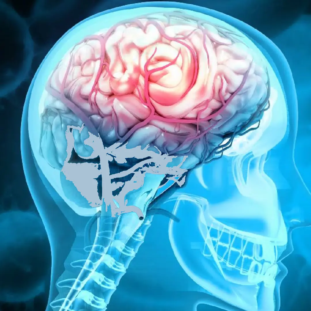
"The extent of surgical resection, assessed according to the Simpson classification, remains the most important predictor of recurrence in meningiomas."
"The surgical indication for asymptomatic meningiomas must be individualised according to age, location, growth rate and surgical risk."
Studies and clinical guidelines underpinning our approach to this condition.
"I arrived with a glioma I had been told was inoperable. Here they reviewed it at the tumour board and offered me a plan. The surgery was a success. Four years on, I am well."
"For years I had a cervical disc herniation, operated on by another team with no improvement. Minimally invasive surgery gave me back my mobility and took the pain away."
"I travelled from outside Spain. The team reviewed my case before I came and gave me a clear plan. Caring people and impeccable technique."
Send us your MRI scan. We review the case at our committee and advise whether surgery is the best option or whether observation is appropriate.