Foraminal stenosis: what it is, symptoms and treatment
Foraminal stenosis is the narrowing of the opening through which each nerve root exits the spine. We explain how it differs from spinal canal stenosis, why it develops, how it is diagnosed and what conservative and surgical treatment options we offer at Brain & Spine Barcelona.
Foraminal stenosis is one of the most common causes of referred pain to the arm or leg (radiculopathy). Understanding its origin and distinguishing it from spinal canal stenosis helps to choose the right treatment, which in most cases does not require surgery.

What is foraminal stenosis?
Between every two vertebrae there is a bony opening called the neural foramen or intervertebral foramen. The nerve root that connects the spinal cord to the rest of the body exits through it. Foraminal stenosis consists of the narrowing of that foramen, which reduces the space available for the nerve root and ends up compressing it.
This compression irritates the nerve and triggers a picture of radiculopathy: pain radiating along the nerve's course, together with tingling, numbness and, in advanced stages, loss of strength. It can occur in the cervical, thoracic or lumbar spine, with the cervical and lumbar regions being the most clinically relevant.
Difference from spinal canal stenosis
Although often confused, they are distinct conditions:
- Spinal canal stenosis (central): narrows the central spinal canal, through which the spinal cord (at cervical and thoracic levels) or the cauda equina (at lumbar level) passes. It typically causes bilateral symptoms or neurogenic claudication (leg pain on walking that improves on sitting).
- Foraminal stenosis (lateral): narrows the foramen through which a specific nerve root exits. It typically produces symptoms of a single nerve root, in one arm or one leg.
Both can coexist in the same patient, and distinguishing them through imaging and examination determines the therapeutic strategy.
Causes of foraminal stenosis
Most cases are degenerative and develop over the years. The most common causes are:
- Facet arthrosis: joint wear generates osteophytes (bony spurs) that invade the foramen.
- Disc herniation: displaced disc material, especially foraminal or extra-foraminal herniation, occupies the root's space.
- Loss of disc height: as the disc dehydrates, the vertebrae approximate and the foramen closes vertically.
- Thickening of the ligamentum flavum.
- Spondylolisthesis: slippage of one vertebra over another deforms the foramen and may cause instability.
- Trauma and fractures causing malalignment.
Symptoms: cervical and lumbar radiculopathy
Cervical foraminal stenosis
When a cervical nerve root is affected, pain starts in the neck and descends through the shoulder and arm to the hand, following a specific pattern. It is accompanied by tingling or numbness in specific fingers, weakness for certain movements and, sometimes, a sense of loss of dexterity. Some neck positions worsen the pain.
Lumbar foraminal stenosis
Involvement of a lumbar nerve root causes sciatica: pain that travels through the buttock and down the back or side of the leg, sometimes to the foot. Tingling, leg weakness and worsening on standing or walking are common, with relief on leaning forward or sitting.
Diagnosis
Diagnosis is clinical and confirmed with complementary tests:
- MRI: the reference test. Shows the compressed root, discs and soft tissues in detail.
- CT: defines the bony component (osteophytes, actual foramen calibre) and is useful when MRI is contraindicated.
- Electromyography (EMG): identifies which nerve root is affected and the degree of compromise, and helps exclude other causes of symptoms.
The imaging has to match the neurological examination: treating the correct nerve root avoids unnecessary surgery.
Conservative treatment
Most patients improve without surgery. Conservative management includes:
- Physiotherapy and therapeutic exercise aimed at decompressing the root and stabilising the spine.
- Analgesia and anti-inflammatory drugs, together with pain neuromodulators when there is a neuropathic component.
- Image-guided epidural or foraminal injections, which reduce inflammation around the root in selected cases.
- Postural habit correction and workplace ergonomics improvement.
Surgical treatment
Surgery is considered when disabling pain persists despite well-conducted conservative treatment, or in the face of progressive neurological deficit (loss of strength). The main options are:
- Foraminotomy: widening of the foramen to decompress the nerve root. It is the technique of choice for isolated foraminal stenosis.
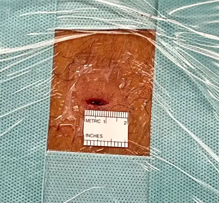
- Minimally invasive and endoscopic decompression: through millimetre-sized incisions, the bone or disc compressing the root is removed, preserving musculature and accelerating recovery.
- Spinal fusion: reserved for cases with associated instability, such as spondylolisthesis, to stabilise the treated segment.
At Brain & Spine Barcelona we always prioritise the least invasive technique that resolves each patient's problem.
Recovery
With minimally invasive and endoscopic techniques many patients are discharged the same day or the following day and resume light activity within a few weeks. When a fusion is performed, consolidation timelines are longer. In all cases, a guided rehabilitation programme improves functional outcome and reduces the risk of recurrence.
Related information
Frequently asked questions
What is foraminal stenosis?
Foraminal stenosis is the narrowing of the intervertebral foramen (neural foramen), the bony opening through which the nerve root exits the spinal column. When this space narrows, the root becomes compressed and pain, tingling or weakness develops in the territory of the affected nerve.
What is the difference between foraminal stenosis and spinal canal stenosis?
Spinal canal stenosis (central stenosis) affects the central spinal canal, through which the spinal cord or cauda equina passes. Foraminal stenosis affects only the lateral foramen through which a specific nerve root exits. This is why foraminal stenosis typically produces single-root symptoms (radiculopathy), whereas central stenosis may cause bilateral symptoms or neurogenic claudication.
What are the symptoms of foraminal stenosis?
Referred pain following the nerve path, tingling, numbness and loss of strength. In the cervical spine it causes neck pain radiating down the arm to the hand; in the lumbar spine it causes sciatica radiating down the leg. Pain typically worsens in positions that close the foramen.
How is it diagnosed?
Diagnosis combines neurological examination with imaging. MRI is the reference test; CT provides detail of the bony component and electromyography (EMG) confirms which root is affected and the degree of compromise.
Does it always need surgery?
No. Most patients improve with conservative treatment: physiotherapy, directed exercise, analgesia and, in selected cases, image-guided epidural or foraminal injections. Surgery is reserved for persistent disabling pain or progressive neurological deficit.
What does surgery for foraminal stenosis involve?
The most common technique is foraminotomy, which widens the foramen to decompress the nerve root, today performed in a minimally invasive or endoscopic manner. If associated instability exists (for example spondylolisthesis), a spinal fusion may be added.
How long is recovery after surgery?
With minimally invasive or endoscopic techniques many patients are discharged the same day or the following day and resume light activity within a few weeks. If a fusion has been performed, recovery timelines are somewhat longer. Guided rehabilitation accelerates recovery.
Where can foraminal stenosis be treated in Barcelona?
The Brain & Spine Barcelona team assesses each case at Hospital El Pilar (Balmes 271, Barcelona) and offers both conservative treatment and minimally invasive, endoscopic and fusion surgery when indicated.
Symptoms of foraminal stenosis?
The Brain & Spine Barcelona team assesses your case and guides you on the most appropriate treatment, conservative or minimally invasive surgery.
Did you find this article useful?
Add us as a preferred source on Google and our articles will show up sooner in your searches.