Dr. Abel Ferrés Pijoan
Neurosurgeon · Spine, neuro-oncology and skull base
View profile →
Gliomas are the most common malignant primary tumour of the central nervous system in adults. The surgical approach aims for maximal safe resection whilst preserving neurological function, combined with radiotherapy and chemotherapy according to histological grade.
Gliomas are tumours that arise from the glial cells of the brain (astrocytes, oligodendrocytes, ependymocytes). They account for approximately 30% of primary brain tumours and 80% of malignant brain tumours. Treatment combines maximal safe resection surgery, radiotherapy and chemotherapy, and its success depends on the histological grade (WHO I-IV) and the extent of resection achieved.
Symptoms depend on tumour location and growth rate. The most common are:
The fundamental oncological principle. Each additional 1% of resection is associated with better survival. We use neuronavigation with preoperative tractography.
Real-time cortical and subcortical mapping for gliomas in eloquent areas (speech, movement). Avoids permanent deficits by modifying the technique according to intraoperative responses.
Continuous motor and somatosensory evoked potentials. Detects any change within fractions of a second to guide surgical adjustments.
5-aminolevulinic acid marks tumour tissue in fluorescent red under blue-light microscopy. Increases the rate of radical resection in glioblastomas.
Immediate confirmation of the extent of resection before closure. Allows further resection if residual tumour is visible.
Neuro-oncology, medical oncology and radiotherapy teams decide on adjuvant treatment after definitive histopathology.
Every patient with a suspected glioma undergoes preoperative multimodal 3 Tesla MRI (spectroscopy, perfusion, tractography). When the tumour contacts eloquent areas, awake surgery is considered after prior neuropsychological assessment. Intraoperative biopsy guides the aggressiveness assessment and allows the surgical strategy to be adjusted. After surgery, the case is presented to the neuro-oncology committee within 7 days to decide on standard adjuvant treatment (radiotherapy plus temozolomide following the Stupp protocol for glioblastomas).
Multimodal brain MRI (functional, DTI, spectroscopy), methionine-PET where indicated, neuropsychological assessment and genetic counselling if suspected. Review by the neuro-oncology committee.
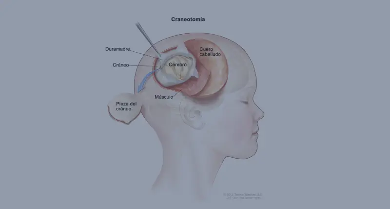
Surgery with neuronavigation, 5-ALA fluorescence for high-grade gliomas, cortical mapping and awake craniotomy when the lesion affects eloquent areas (language, motor). Intraoperative MRI where available.
ICU for 24 hours, postoperative MRI within 72 hours to measure the extent of resection. Chemoradiotherapy following the Stupp protocol begins 3-4 weeks later for high-grade gliomas.
Typical hospital stay of 4-7 days. Personalised neurocognitive rehabilitation and multidisciplinary committee follow-up every 3 months.
Prognosis depends on histological grade: low-grade gliomas (WHO II) have median survival of 7-10 years, whilst glioblastoma (WHO IV) has a median of 14-18 months with standard treatment. Resection of 98% or more is associated with a significant improvement in overall and progression-free survival. Typical postoperative recovery allows discharge in 4-6 days.
Regarding functional recovery, approximately 70% of patients recover or improve their neurological level during the first 4-8 weeks after surgery. Beyond that period, spontaneous improvement is very limited: only around 50% experience further improvement without active treatment. This is why early surgical indication and intraoperative mapping are decisive: in oncological neurosurgery, waiting is not a therapeutic option.
"Maximal safe resection remains the most important modifiable prognostic factor in high-grade gliomas. Techniques such as awake surgery and 5-ALA have significantly increased rates of radical resection."
"In low-grade gliomas, the extent of resection is associated with improvement in overall and progression-free survival."
Studies and clinical guidelines underpinning our approach to this condition.
"I arrived with a glioma I had been told was inoperable. Here they reviewed it at the tumour board and offered me a plan. The surgery was a success. Four years on, I am well."
"For years I had a cervical disc herniation, operated on by another team with no improvement. Minimally invasive surgery gave me back my mobility and took the pain away."
"I travelled from outside Spain. The team reviewed my case before I came and gave me a clear plan. Caring people and impeccable technique."
Send us your MRI scan and histopathology report if available. We review your case at our neuro-oncology committee and respond within 24-48 hours.