Brain tumour diagnosis and treatment in Barcelona
A clear clinical guide to brain tumours: what they are, how they are diagnosed with MRI and biopsy, and what treatment options (awake craniotomy, radiotherapy and multidisciplinary tumour board) advanced neurosurgery offers in Barcelona.
A brain tumour diagnosis understandably causes great anxiety, but modern neuro-oncology allows precise confirmation of the tumour type and the design of an individualised treatment plan. In Barcelona, the combination of advanced neuroimaging, molecular analysis, high-technology surgery and multidisciplinary tumour boards offers each patient the best possible strategy.
What is a brain tumour?
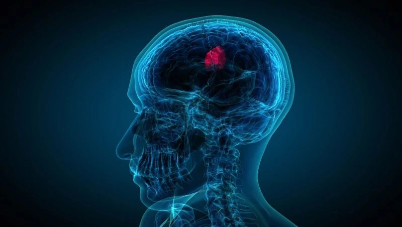
A brain tumour is an abnormal growth of cells inside the brain or in the structures surrounding it (meninges, cranial nerves, pituitary gland). Not all brain tumours are cancer: there are benign tumours, slow-growing and well-defined, and malignant tumours, more aggressive and infiltrative. It is worth noting that even a benign tumour can cause significant symptoms if it compresses important structures depending on its location.
Two main groups are distinguished: primary tumours, which arise from neural tissue itself or its coverings, and metastatic or secondary tumours, which originate in a cancer from another organ (lung, breast, kidney, melanoma...) that has spread to the brain.
Types of brain tumours
Knowing the specific tumour type is fundamental, as it determines prognosis and treatment. The most common are:
- Gliomas: arise from glial cells. They include astrocytomas, oligodendrogliomas and, in their most aggressive form, glioblastoma. Their grade (I to IV) and molecular profile guide treatment.
- Meningiomas: arise from the meninges. Usually benign and slow-growing; many are cured by surgery and some are monitored without immediate intervention.
- Brain metastases: the most common cause of brain tumour in adults, arising from cancers in other organs.
- Pituitary adenomas: pituitary tumours that may affect hormones or vision.
- Schwannomas (neuromas): such as the vestibular schwannoma, affecting hearing and balance.
- Others: medulloblastomas, ependymomas, skull base tumours and tumours specific to childhood.
The distinction between benign and malignant is not the only factor: location, size and, above all, molecular analysis have transformed the modern classification of brain tumours.
Warning symptoms
Symptoms depend on the tumour's location and size. Specialist consultation is warranted for persistent or recently onset neurological signs:
- New, severe and progressive headache, often worse in the morning or waking the patient at night.
- Nausea and vomiting without a clear digestive cause.
- Epileptic seizures (convulsions), especially in an adult with no prior history.
- Loss of strength, tingling or loss of sensation in part of the body.
- Disturbances of vision, speech, balance or gait.
- Changes in personality, memory, concentration or behaviour.
These symptoms do not necessarily indicate a tumour (they are non-specific), but they justify specialist assessment when persistent or progressive.
Diagnosis of brain tumours
Diagnosis begins with a neurological examination and is supported by neuroimaging. MRI with and without contrast is the reference test: it shows the location, size and characteristics of the tumour. It is supplemented by advanced techniques that help characterise it and plan surgery safely:
- MR spectroscopy: analyses the chemical composition of the tissue and indicates the nature of the tumour.
- Perfusion MRI: evaluates vascularisation and aggressiveness.
- Functional MRI and tractography: localise language and movement areas and nerve tracts to preserve them during surgery.
A definitive diagnosis requires, in most cases, a biopsy or analysis of the sample obtained during surgery. Histopathological study and, increasingly, molecular and genetic analysis confirm the exact tumour type and enable precision medicine.
Treatment of brain tumours
Treatment is always decided individually in a neuro-oncology multidisciplinary tumour board, where neurosurgeons, oncologists, radiation oncologists, neuroradiologists and pathologists agree on the best strategy. The main tools are:
Surgery
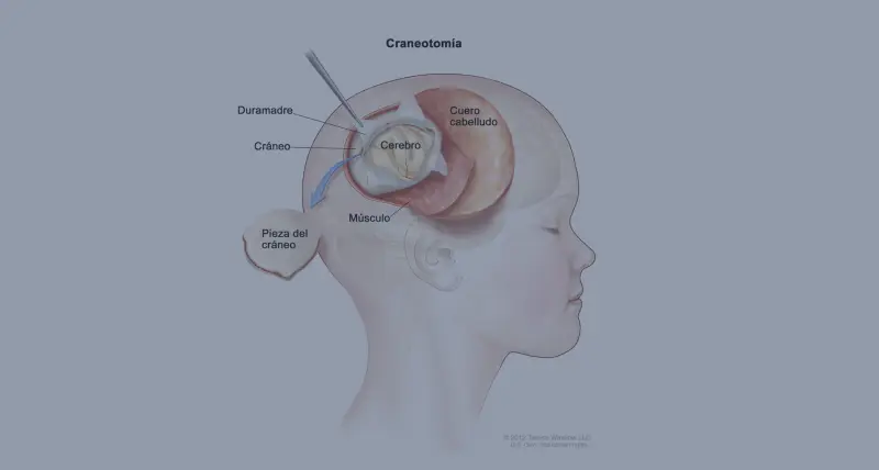
Surgery is, in many cases, the first step. The goal is to remove as much tumour as possible whilst preserving neurological function. For tumours near eloquent areas, awake craniotomy with cortical mapping is used: the patient cooperates by speaking or moving whilst the team identifies and respects critical zones. Microsurgery, neuronavigation and intraoperative neurophysiological monitoring increase precision and safety. You can find further details about our brain tumour surgery and the full range of brain surgery procedures.
Radiotherapy and chemotherapy
Radiotherapy (including stereotactic radiosurgery for small or multiple lesions) and chemotherapy or targeted therapies complement or replace surgery depending on tumour type. In many malignant tumours several modalities are combined to improve disease control.
Minimally invasive surgery and technology
Modern neurosurgery tends towards minimally invasive techniques that reduce trauma, complications and recovery times. Endoscopic neurosurgery, for example the endoscopic endonasal approach to skull base and pituitary tumours, allows access to deep lesions through natural corridors. Neuronavigation, intraoperative fluorescence and neurophysiological monitoring allow more tumour to be safely removed.
Second opinion
Facing such a delicate diagnosis, a neurosurgical second opinion provides reassurance and, on occasion, opens new treatment possibilities. It allows review of imaging, confirmation of the diagnosis and assessment of all alternatives, especially when surgery has been ruled out at another centre or for complex cases. You can find out about our specialists' background on the medical team page.
Prognosis
The prognosis of a brain tumour varies enormously depending on type, grade, location, extent of resection and response to treatment. Many benign tumours are cured by surgery. For malignant tumours, advances in surgery, radiotherapy, targeted therapies and the molecular approach have significantly improved survival and quality of life in recent years. Early diagnosis and assessment by an expert team are therefore decisive.
Frequently asked questions
Are all brain tumours cancer?
No. There are benign (non-cancerous) brain tumours, such as many meningiomas, and malignant tumours, such as high-grade gliomas or metastases. Even a benign tumour can cause significant symptoms depending on its size and location, so every case must be assessed individually.
What symptoms should raise suspicion of a brain tumour?
New, severe and progressive headache (often worse in the morning), nausea or vomiting, epileptic seizures, changes in vision, loss of strength or sensation, speech, balance or behavioural disturbances. Persistent neurological symptoms warrant specialist assessment.
What tests are used to diagnose a brain tumour?
MRI with and without contrast is the reference test. It is supplemented by advanced techniques such as spectroscopy, perfusion imaging, functional MRI and tractography, and by biopsy or molecular analysis of the sample to confirm the diagnosis and guide treatment.
What does awake craniotomy involve?
It is a technique for tumours near eloquent areas (language, movement). The patient cooperates during part of the procedure while cortical mapping is performed, allowing maximum tumour removal whilst preserving essential functions.
Is surgery always necessary?
Not always. The plan depends on the tumour type, its location, the patient's condition and molecular analysis. In some cases radiotherapy, chemotherapy or active surveillance is prioritised. Decisions are made in a multidisciplinary tumour board.
Is it worth seeking a second opinion?
Yes. A neurosurgical second opinion allows confirmation of diagnosis, review of imaging and assessment of all treatment options. It is especially useful when surgery has been ruled out elsewhere or for complex diagnoses.
What is a neuro-oncology multidisciplinary team?
It is a meeting of neurosurgeons, oncologists, radiation oncologists, neuroradiologists and pathologists who jointly study each case to define the best individualised treatment strategy.
Is the prognosis always poor?
No. Prognosis varies enormously depending on tumour type, grade, location and response to treatment. Many benign tumours are cured by surgery, and advances in neuro-oncology have significantly improved survival and quality of life for many malignant tumours.
Need to assess a brain tumour or seek a second opinion?
The Brain & Spine Barcelona team can review your case and imaging, and guide you on the most appropriate diagnostic and treatment options.